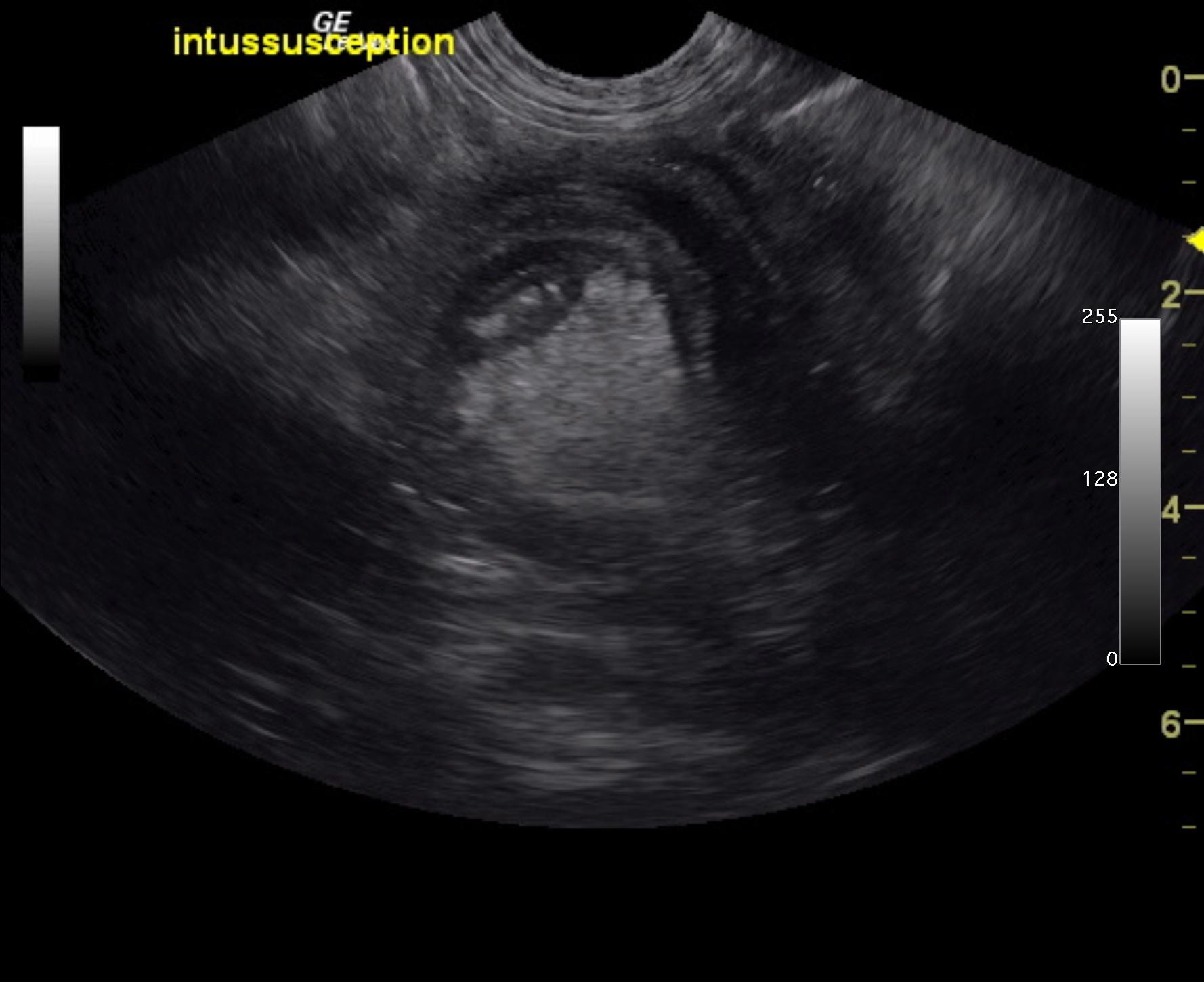
A 6-month-old intact male Labrador was presented for evaluation of diarrhea and vomiting for approximately 1 week and more recently only hemorrhagic diarrhea.
A 6-month-old intact male Labrador was presented for evaluation of diarrhea and vomiting for approximately 1 week and more recently only hemorrhagic diarrhea.