The patient is a canine Mix, NM, 11 years, 19.8 lbs. Clinical exam findings: decreased appetite, weight loss, febrile. Albumin 2.2, amylase 1633, hematocrit 27, white count 47000 with neutrophilia and band cells. Urine specific gravity 1.035, pH 6
The patient is a canine Mix, NM, 11 years, 19.8 lbs. Clinical exam findings: decreased appetite, weight loss, febrile. Albumin 2.2, amylase 1633, hematocrit 27, white count 47000 with neutrophilia and band cells. Urine specific gravity 1.035, pH 6
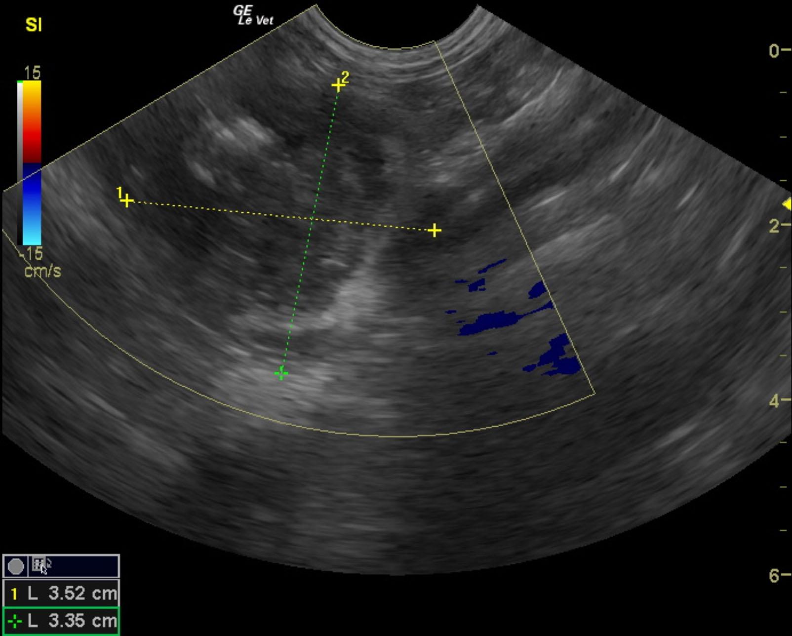
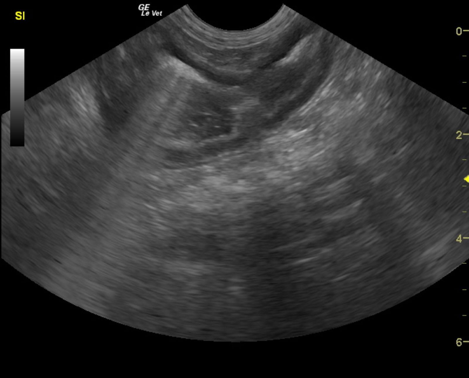
Mid to distal small intestinal mass.
Regional lymph node enlargement.
The intestinal mass appears resectable. However, given the lymph node enlargement, both differentials of reactive or infiltrative lymph node would need to be evaluated. Lymph node removal at the time of surgery is recommended. Alternatively, ultrasound-guided FNA of the mass and lymph node could be performed for further definition.
The gastrointestinal tract revealed a complex 3.52 cm small intestinal mass obliterating mural detail. In this area there was regional inflammation. Regional lymph nodes were enlarged and measured 1.5 cm each with pericapsular inflammatory pattern. This was adjacent to the jejunum.
None
PLE, PLN, pancreatitis, gastroenteritis, neoplasia, infectious.
FNA of mass and LN: results unavailable