A 1-year-old female cat was presented for evaluation of chronic diarrhea and weight loss that had not responded to metronidazole and deworming. Urinalysis, CBC, serum biochemistry, and T4 were all within reference range.
A 1-year-old female cat was presented for evaluation of chronic diarrhea and weight loss that had not responded to metronidazole and deworming. Urinalysis, CBC, serum biochemistry, and T4 were all within reference range.
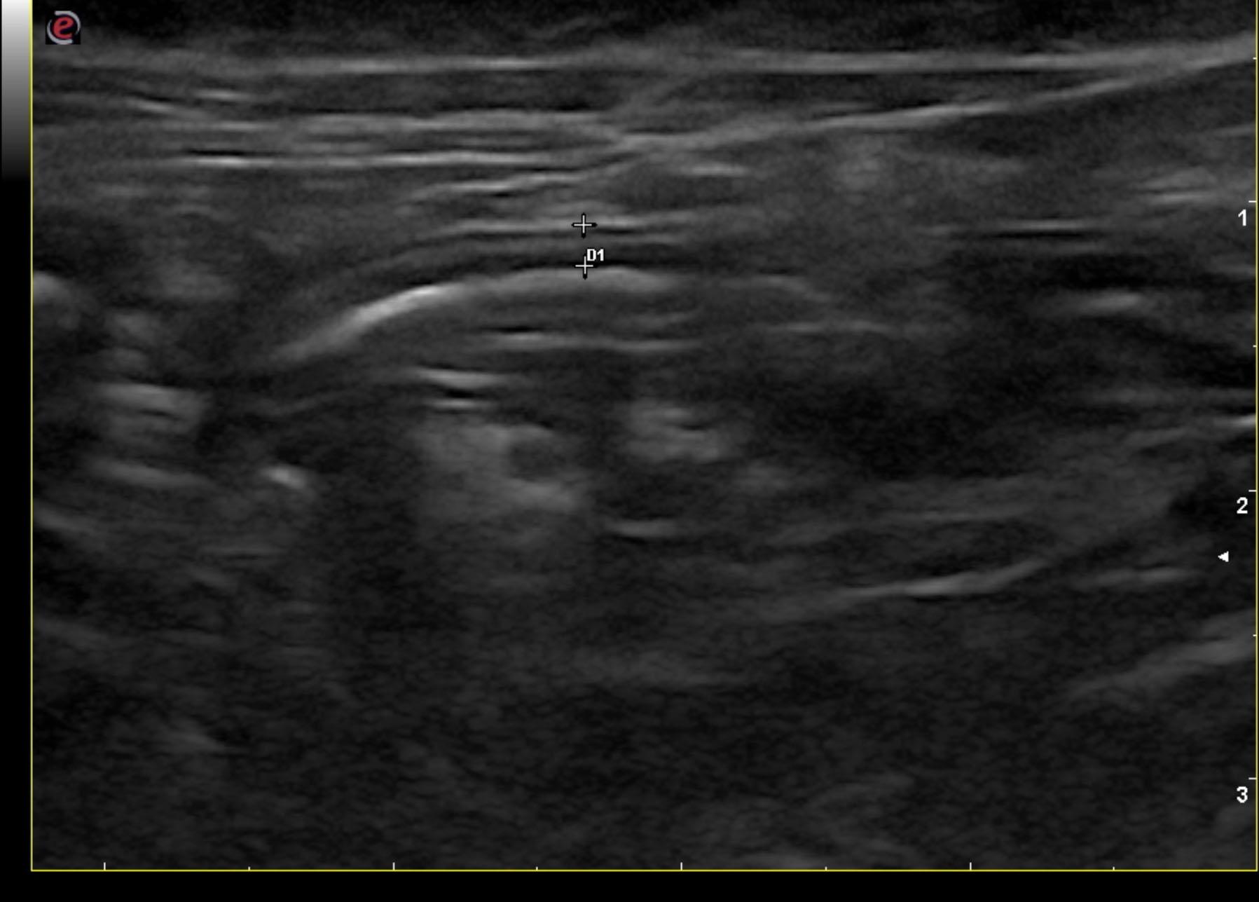
Stricturing colonic lesion with regional inflammation and localized peritonitis. Ultrasound-guided FNA of the colic lymph node was performed without complication. Subtotal colectomy is strongly recommended in this patient with regional lymph node removal. There was no overt evidence of metastatic disease.
The gastrointestinal presentation revealed areas of “ropey” small intestinal wall with slight disruption of the normal 1:3 muscularis/mucosal ratio. The intestinal mucosa was slightly irregular, thickened and hyperechoic suggestive of low grade, chronic inflammation. Hard stool was noted in the colon with focal, descending colonic thickening and regional lymphadenopathy. This appears to be partially stricturing. Focal mineralization was noted within the colonic wall. The stricturing is at the level of the body of the urinary bladder.
None
IBD, dietary hypersensitivity, granulomatous enteritis, lymphoma
Ultrasound-guided FNA of the colic lymph node