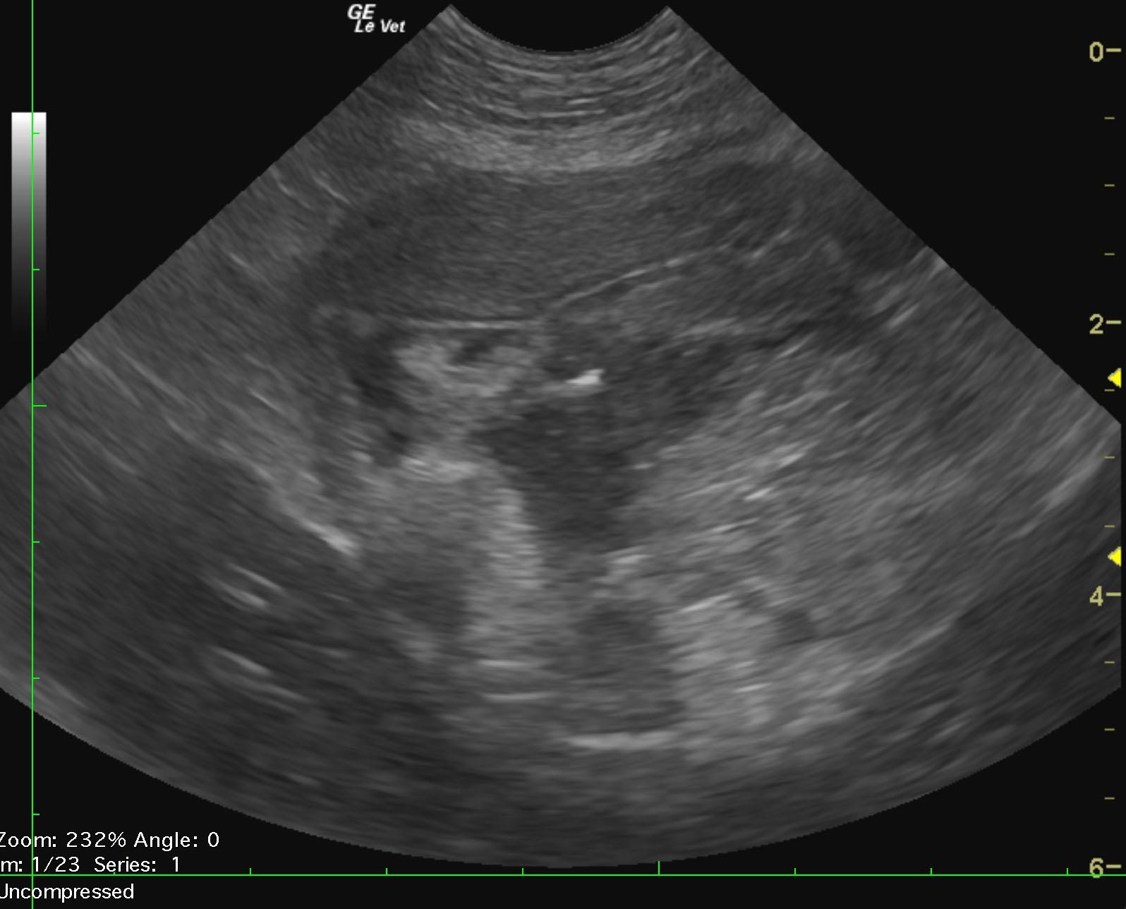
The patient, a 10 year old FS Maltese, was presented for vomiting and diarrhea. On blood work she was found to have minor anemia and a WBC of 33,000.
The patient, a 10 year old FS Maltese, was presented for vomiting and diarrhea. On blood work she was found to have minor anemia and a WBC of 33,000.