An 11-year-old SF mixed breed dog was presented for evaluation of unresponsive UTI with abdominal radiographs showing possible unilateral renomegaly.
An 11-year-old SF mixed breed dog was presented for evaluation of unresponsive UTI with abdominal radiographs showing possible unilateral renomegaly.
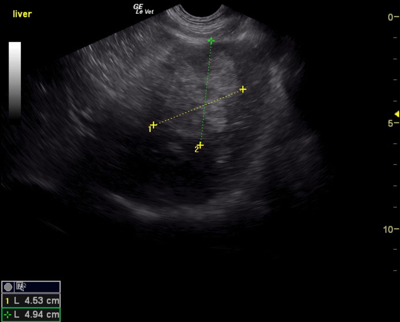
Left sided liver mass.
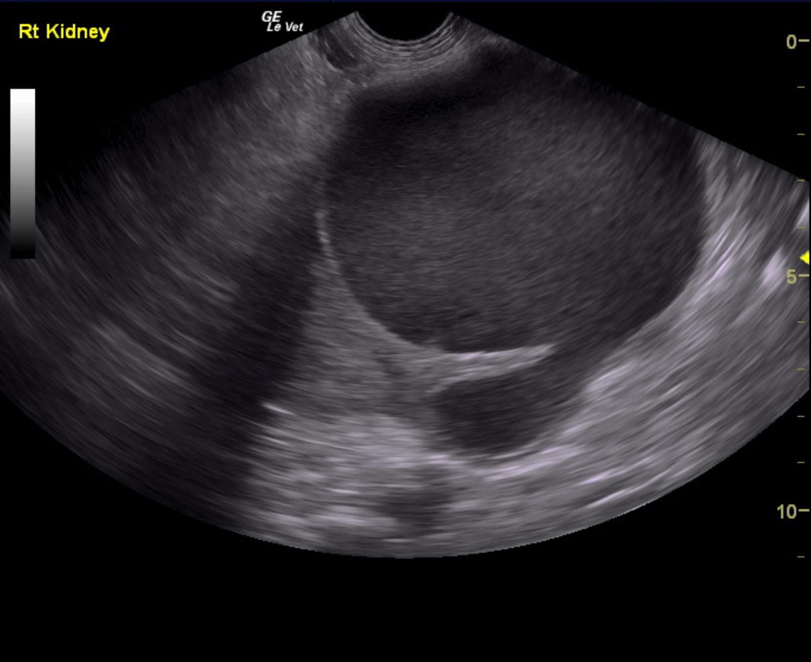
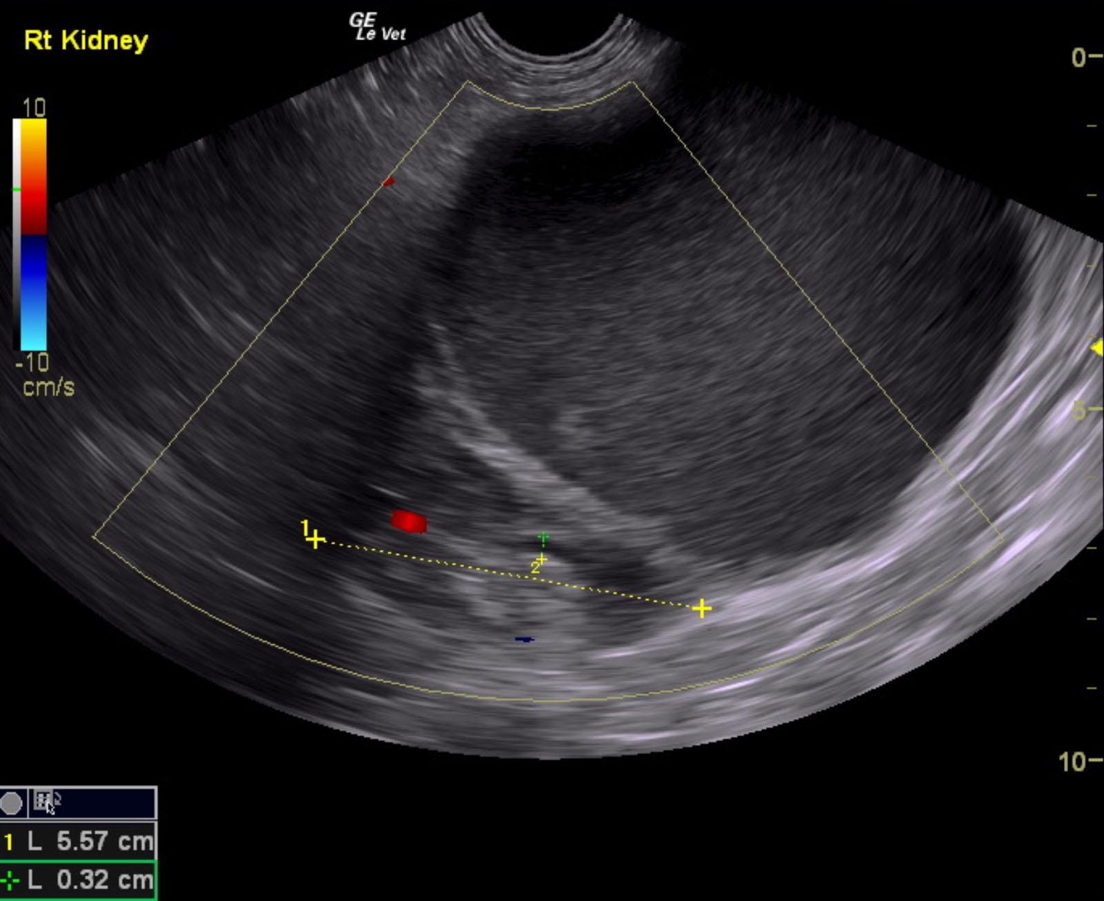
Echogenic renal cyst, potential underlying neoplasia. Pyelectasia in the right kidney, consistent with concurrent pyelonephritis.
Right nephrectomy is recommended along with left liver lobectomy. Pericapsular inflammatory pattern was also noted around the cystic portion of the right kidney. This is suggestive for infection or potential emerging neoplasia.
The liver presented a mixed, echogenic and cystic mass that was deriving from the mid left liver. This is strongly consistent with hepatic carcinoma and appears resectable with left lobectomy. The mass measured 4.9 x 4.5 cm.
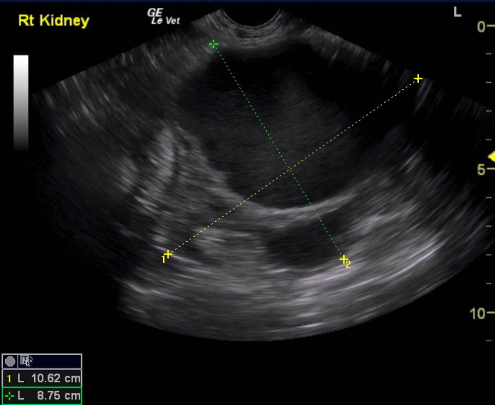
The left kidney was uniform with no evident pathology and measured 5.93 cm. The right kidney in this patient presented large, echogenic cysts that measured approximately 8.26 cm and was deriving from the renal cortex. Pyelectasia of the right kidney was also noted and measured 0.41 cm. The right kidney itself measured 5.57 cm. The kidney along with the cyst measured 10.62 x 8.75 cm.
None
Hydronephrosis, pyelonephritis, neoplasia, renolith, renal cysts, renal abscess
None