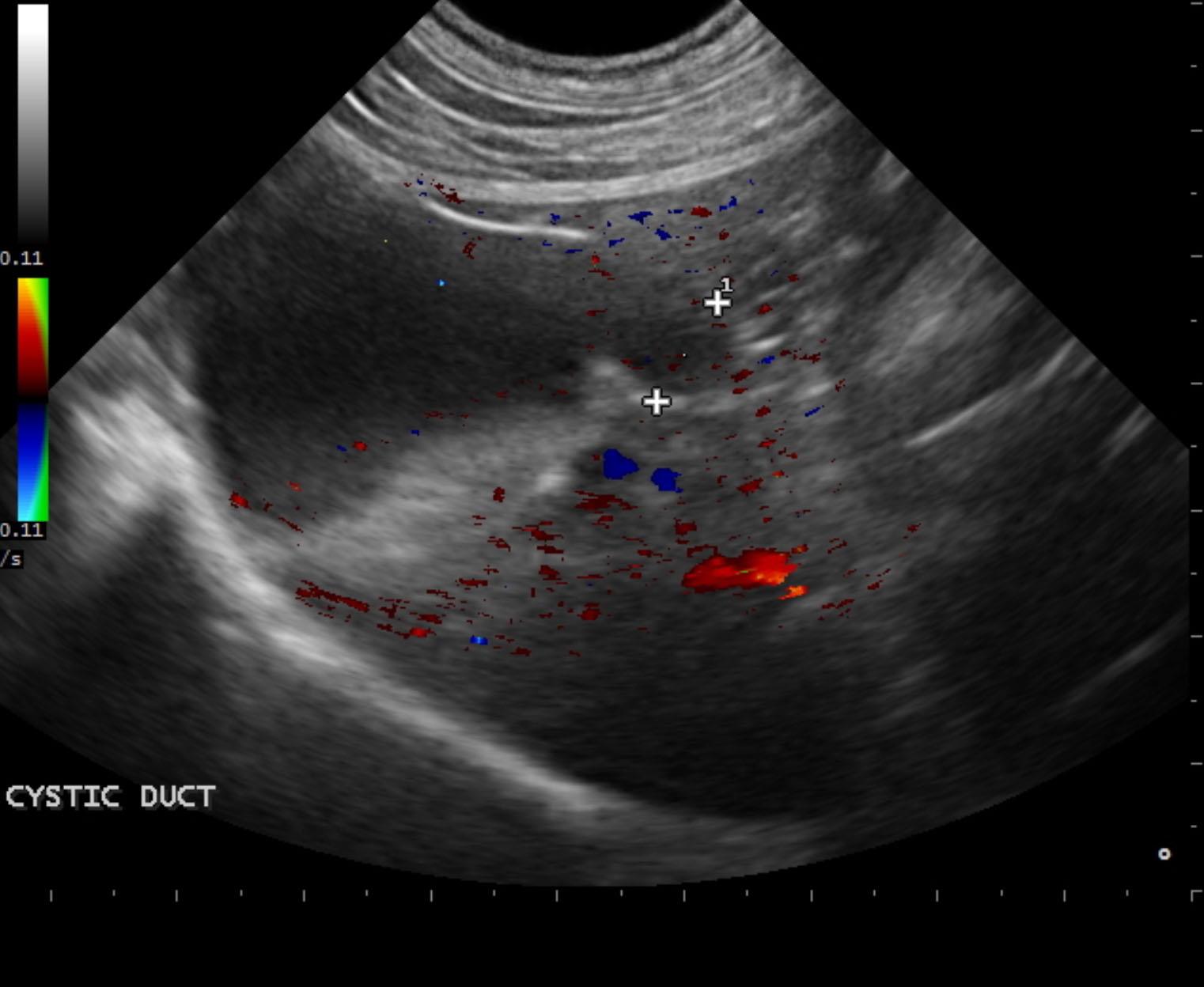
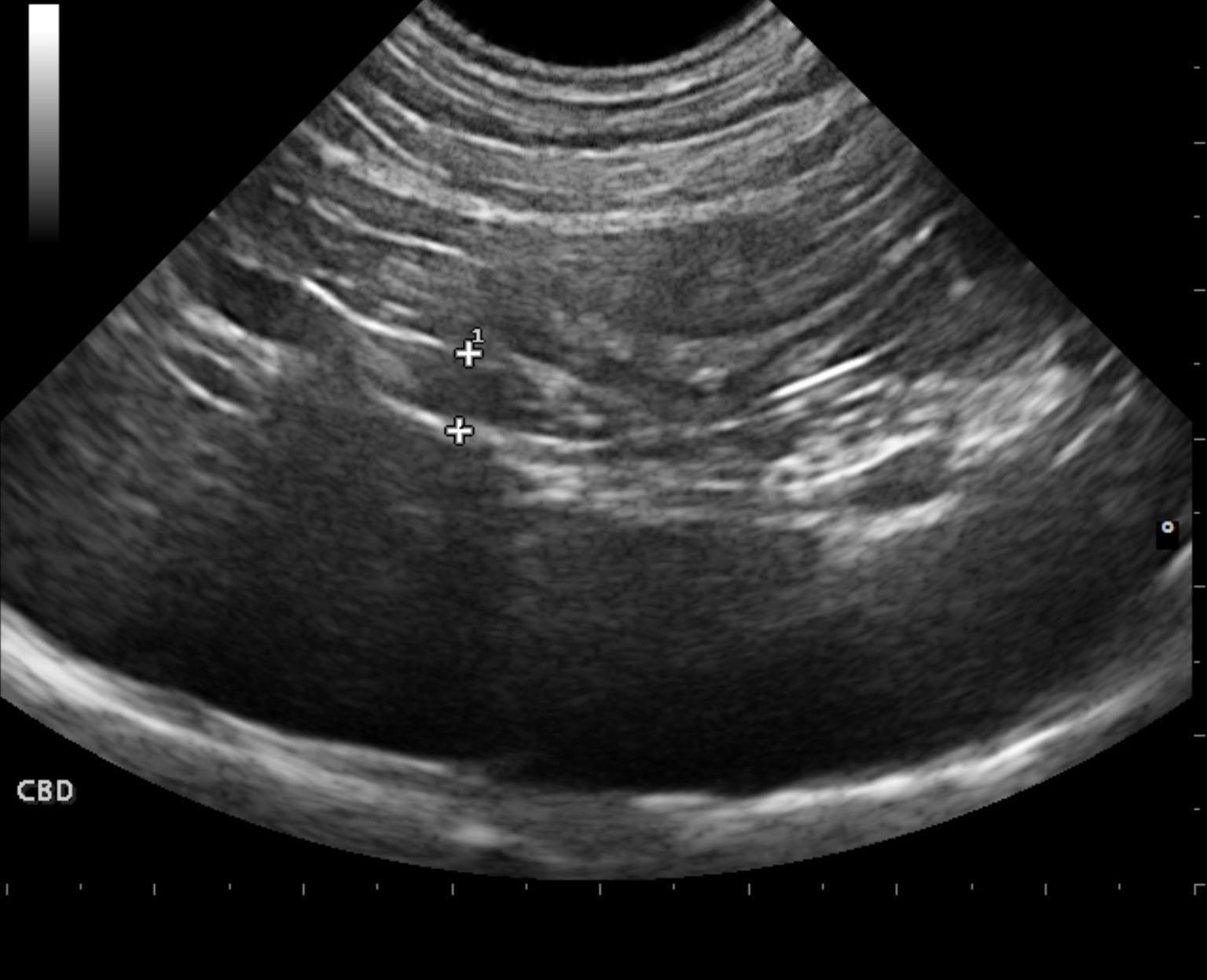
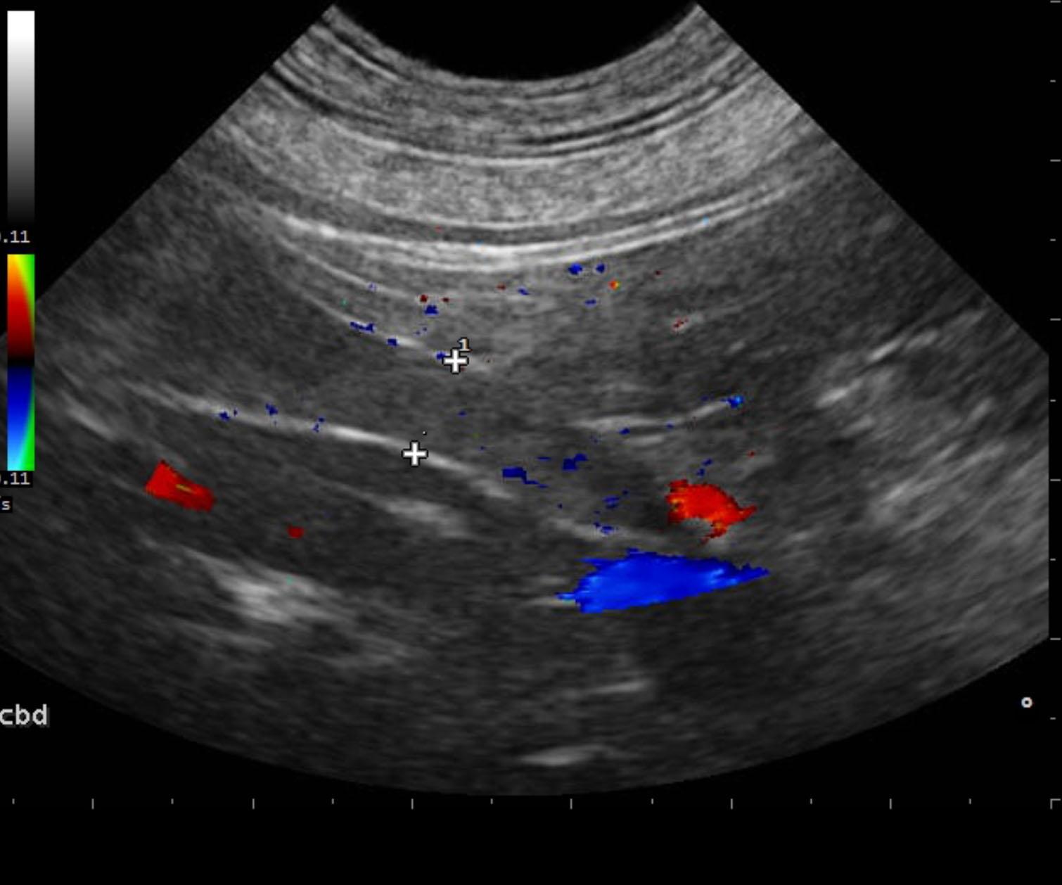
Posthepatic obstruction with likely mucoduct and congested gallbladder.
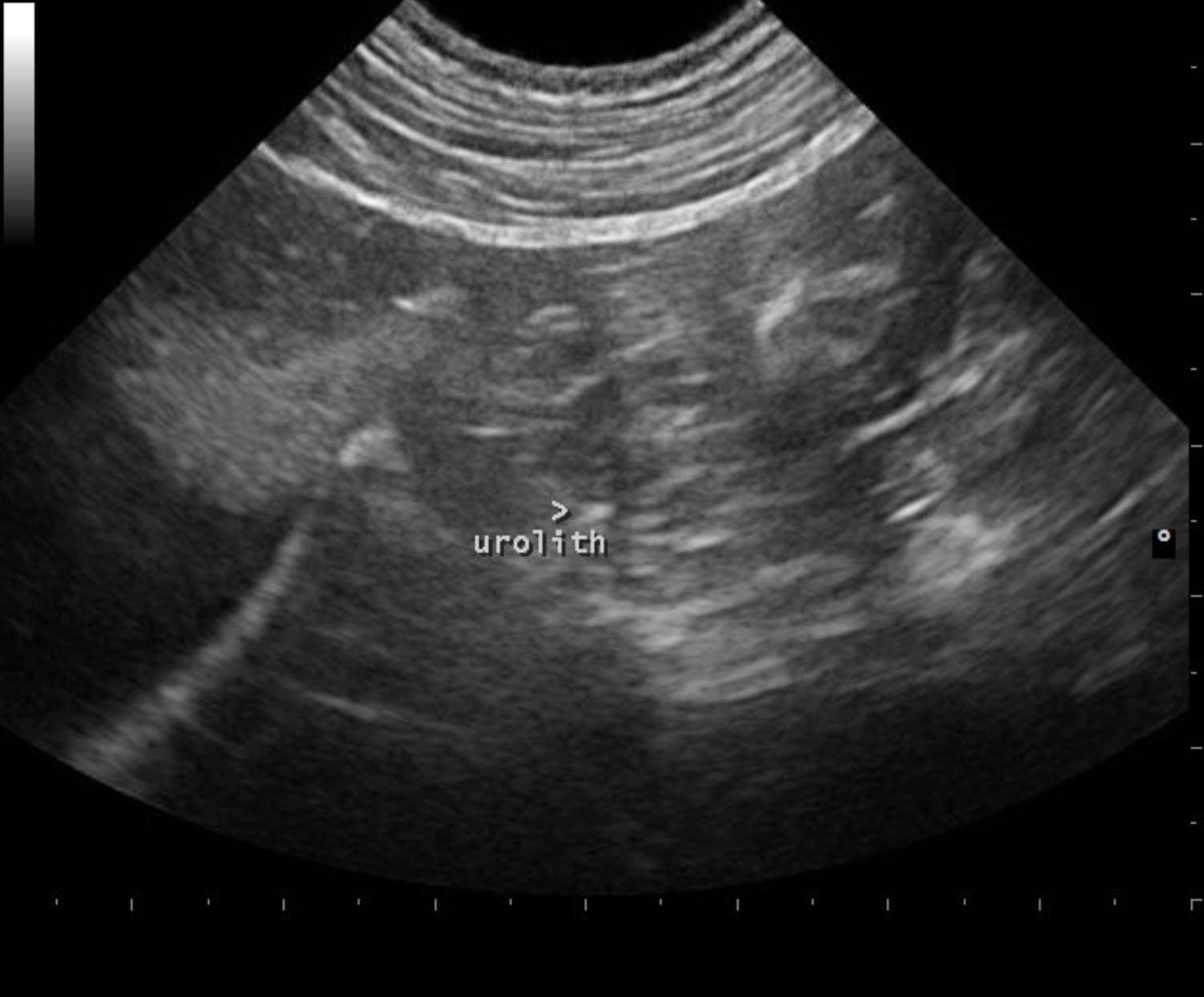
A small calculus, labeled urolith, was noted in the duodenal papilla in this patient, yet this is not likely the complete issue regarding the common bile duct obstruction. If the patient is stable, then medical therapy could be attempted and recheck sonogram in 48 to 72 hours; however, surgical intervention will likely be necessary in this case. Hypercoagulable state should be considered. Leptospirosis should also be considered to be complete in this patient; however, ampicillin and metronidazole plus Ursodiol therapy could be attempted medically; however surgical intervention with bile duct lavage plus or minus cholecystoduodenostomy will likely be the best option in this patient.