A 3-year-old SF Chihuahua mix was presented for evaluation of weight loss and elevated ALT activity (382-390).
A 3-year-old SF Chihuahua mix was presented for evaluation of weight loss and elevated ALT activity (382-390).
Structurally unremarkable inflammatory hepatopathy.
There was no structural evidence of disease.
I recommend treatment based on cytology. If lymphoplasmacytic predominance is noted in the inflammatory pattern then antigen surveillance protocol would be recommended. There was no structural evidence of disease that would be responsible for the weight loss.
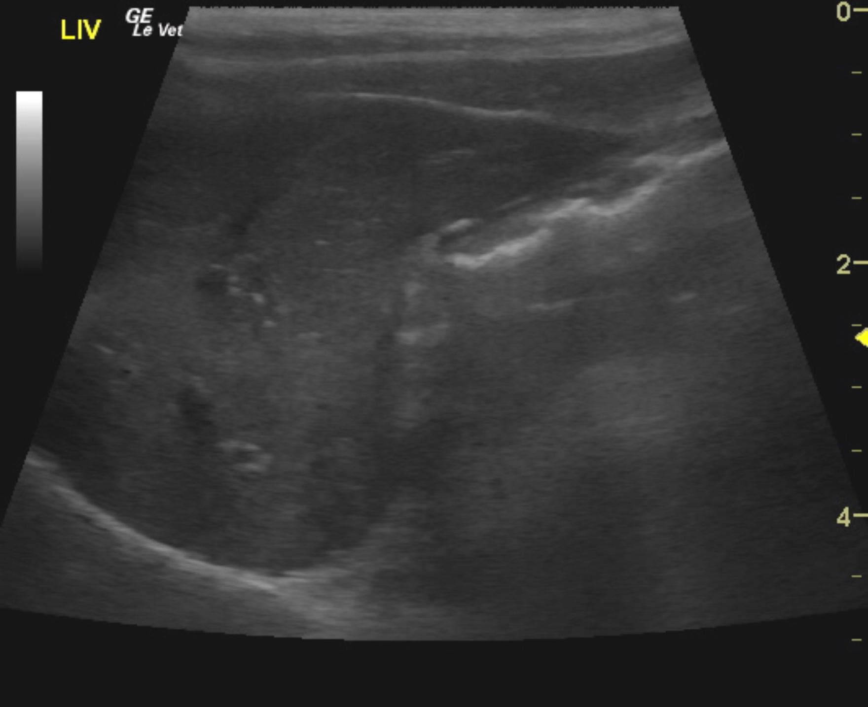
The liver images from right and left intercostal as well as subcostal views revealed subjectively normal liver size, contour, and structure. Parenchymal echogenicity was naturally coarse and hypoechoic to the spleen. Vascular and biliary tracts were of normal volume and no evidence of congestion was noted. The portal vein to vena cava ratio was 1:1 each measuring 0.5 cm. The gallbladder presented thin walls with normal, primarily anechoic content. The cystic and common bile ducts were normal. No pathological hepatic lymphadenopathy was evident. No overt structural evidence of inflammatory, infiltrative or regenerative pathology was noted. Ultrasound-guided FNA of the liver was performed without complication.
None
Vacuolar hepatopathy, reactive hepatopathy, acute hepatitis (viral, bacterial, toxins), drug-induced, infiltrative neoplasia (lymphoma, mast cell tumor)
FNA of the liver revealed mild to moderate glycogen-type hepatocellular vacuolar change. Possible mild small lymphocytosis.