03_00452 Ceaser B Splenoazygos shunt

Case Study

03_00452 Ceaser B Splenoazygos shunt

A 6-year-old NM Silky Terrier was presented for evaluation of lethargy and vomiting and was hospitalized for IV fluids and medications. Post-prandial bile acids were elevated (147).


Sonographic Differential Diagnosis

Splenoazygos shunt

Image Interpretation

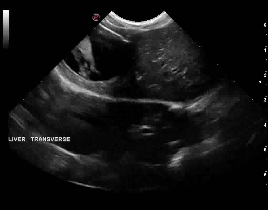
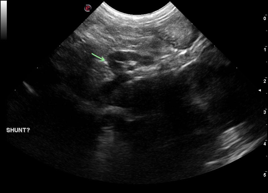
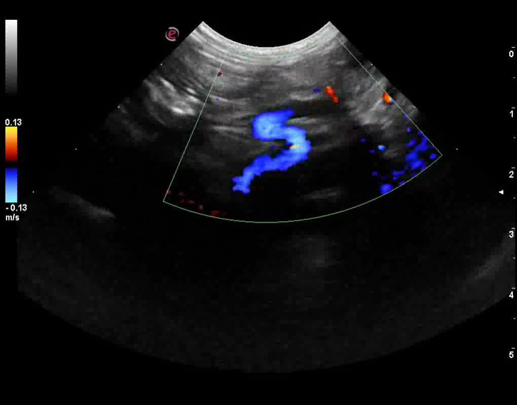
Dorsally directed erroneous vessel entering into the aortic hiatus consistent with splenoazygos shunt. Microhepatica likely owing to portal hypoplasia and/or EHPSS.
DX

Splenoazygos shunt

Outcome

None

Comments

Case courtesy of Marty Henderson, SonoVet, Inc.

Clinical Differential Diagnosis

Liver – chronic hepatopathy, cirrhosis, neoplasia, granulomatous disease
Porto-caval shunt, primary portal vein hypoplasia

Sampling

None

Patient Information

Gender : Male, Neutered
Species : Canine
Type of Imaging : Ultrasound
Status : Complete

Clinical Signs

  • Lethargy
  • Vomiting

Images

3_18_00000040793_copyvlcsnap-2015-08-01-13h00m12s227vlcsnap-2015-08-01-13h01m12s213

Clinical Signs

  • Lethargy
  • Vomiting