03_00445 Oliver V FIP, wet form

Case Study

03_00445 Oliver V FIP, wet form

 A 16-week-old intact male Siamese was presented for evaluation of ascites. Abnormalities on CBC and serum biochemistry were non-regenerative anemia, hypoalbuminemia, elevated bilirubin, and PCR positive for Corona virus. The ascites was quantified as a non-septic exudate.

DX

FIP, wet form

Sonographic Differential Diagnosis

Potential granulomatous, hepatic changes.
Strong suspicion for wet form FIP.
Sludging of blood in the portal vein and vena cava. PCR evaluation of the fluid for FIP is recommended. Poor prognosis.

Image Interpretation

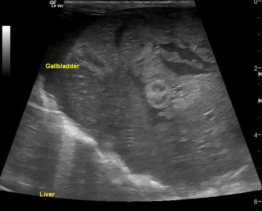
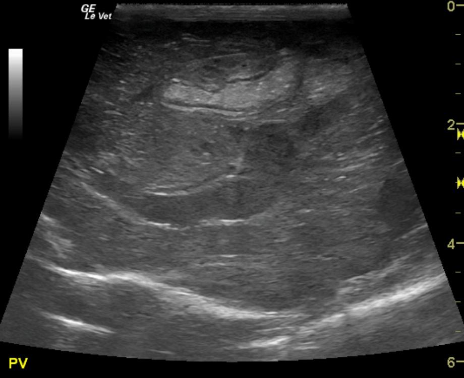
The liver was mildly heterogenous with increased portal markings and coarse architecture. The portal vein was dilated with echogenic debris. However, no evidence for portal hypertension was noted. The portal vein velocity was normal at 25 cm/sec. The echogenic debris within the portal vein was also repeated in the vena cava. This is suggestive for inspissation and a high predisposition for clot formation. The common bile duct measured 0.3 cm. The gallbladder was collapsed and double layered with debris.
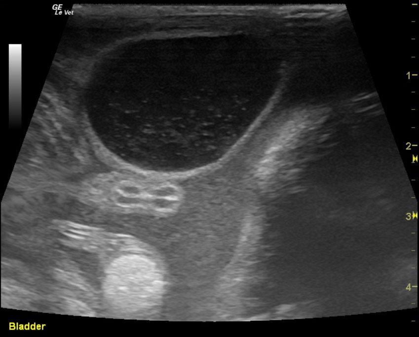
The abdomen in this patient presented a large amount of echogenic free fluid.

Outcome

None

Clinical Differential Diagnosis

Exudative FIP, hepatopathy

Sampling

None

Patient Information

Gender : Male, Intact
Species : Feline
Type of Imaging : Ultrasound
Status : Complete

Clinical Signs

  • Ascites

Exam Finding

  • Ascites

Images

livgbpvunnamed

Blood Chemistry

  • Albumin, Low
  • Total Bilirubin, High

CBC

  • RBC, Low

Clinical Signs

  • Ascites
Skip to content