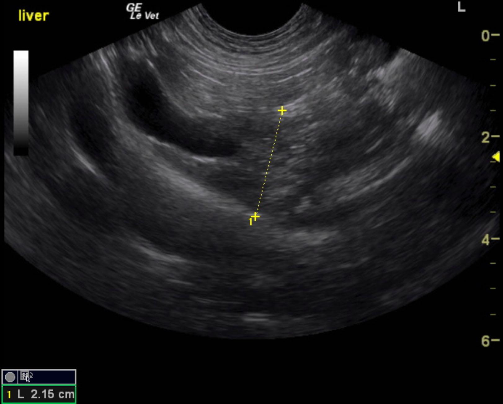
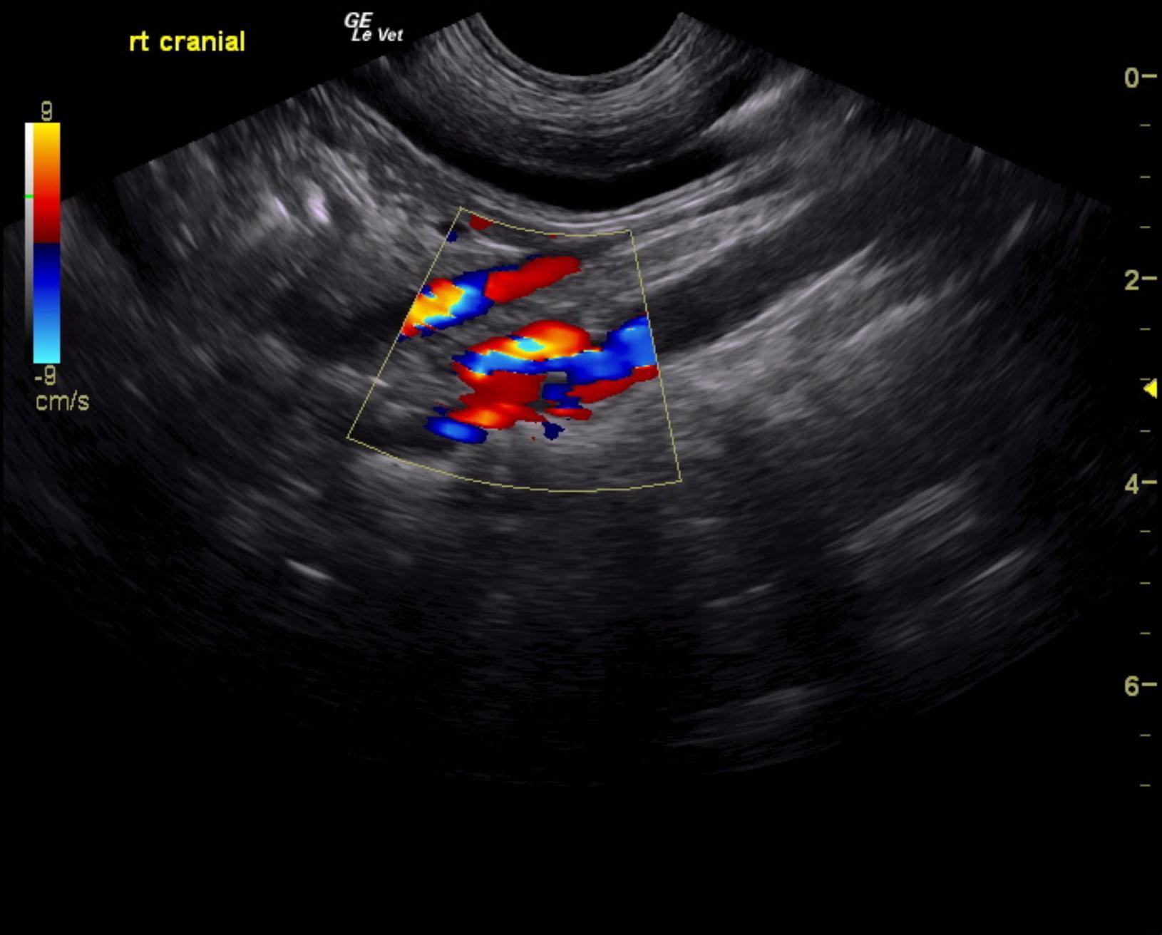
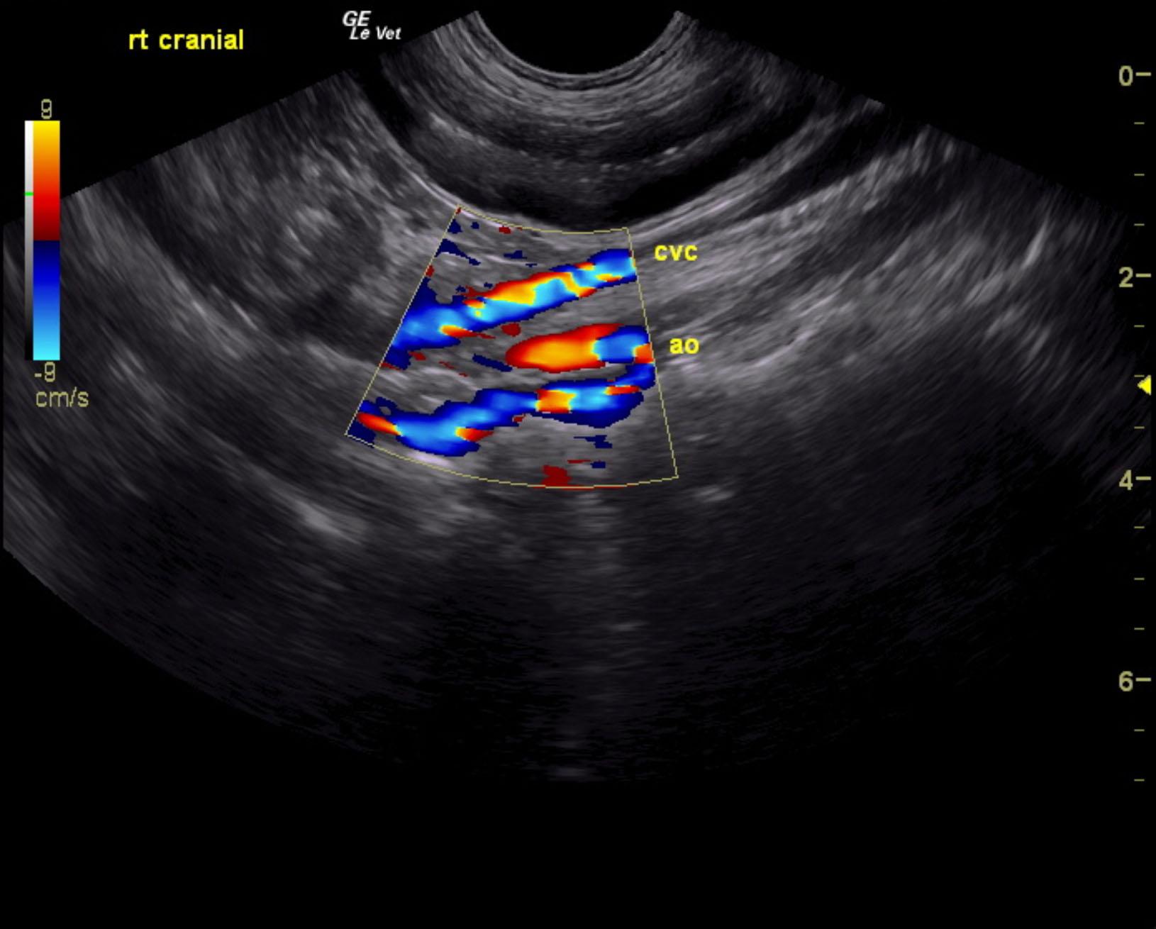
The liver was significantly subnormal in size. The width in short axis measured 2.1 cm. Increased portal markings were noted. Double aorta sign was noted and measured 0.5 cm. This would be consistent with azygos shunt. The vena cava to aortic ratio was normal at 0.3 cm. Significant gas artifact was noted in the region of the portal hilus. An extrahepatic portosystemic shunt was clearly visible entering into the azygos region. This measured approximately 0.6 cm in width. This was in the position of a splenoazygos shunt. However, gastroazygos shunt could not be entirely ruled out. The dorsal aspect of the shunt was best represented in video #1 as entrance into the aortic hiatus of the shunt was clearly visible. The gallbladder was unremarkable.
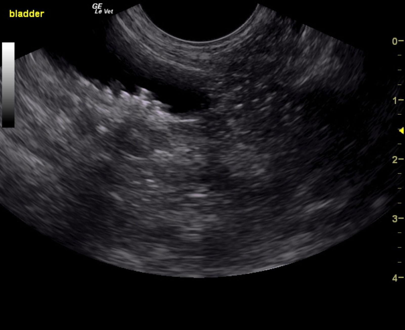
The urinary bladder, trigone and pelvic urethra presented normal wall thicknesses with anechoic urine and normal tone. Bladder calculi were noted with an accumulation of which measuring 1.06 cm. No evidence of inflammatory or neoplastic changes were noted. The ureters were not visible and considered normal.