A 7-year-old NM terrier mixed canine was presented with a 3 day history of vomiting and anorexia that initially improved with fluids, lactulose and clavamox but there was vomiting, lethargy and diarrhea for the past 24 hours. Initial bloods showed left shift neutrophilia, hypoalbuminemia, elevated liver enzyme activity, bilirubinemia, and hypoglycemia. On follow up bloods, progressively elevated ALP activity with normalization of ALT, bilirubin, albumin, and neutrophils was evident
A 7-year-old NM terrier mixed canine was presented with a 3 day history of vomiting and anorexia that initially improved with fluids, lactulose and clavamox but there was vomiting, lethargy and diarrhea for the past 24 hours. Initial bloods showed left shift neutrophilia, hypoalbuminemia, elevated liver enzyme activity, bilirubinemia, and hypoglycemia. On follow up bloods, progressively elevated ALP activity with normalization of ALT, bilirubin, albumin, and neutrophils was evident
Case Study
03_00231 Whiley S Mucocele
Sonographic Differential Diagnosis
The most likely etiology for the gall bladder pathology would be a mucocele with cholicystitis another possibility. This would explain the progressive elevation of ALP activity with normalization of ALT, bilirubin, and albumin. As there is a weak association between mucocele and Cushing’s disease, screening for Cushing’s disease may be indicated if there are supporting clinical signs. Recommended therapy is surgery (cholecystectomy, cholecystotomy, cholecysto-enterostomy) as non-surgical resolution has only been reported in a hand-full of cases. Non-surgical management would include liver diet, antibiotics (penicillin, cephalosporin), and anti-emetics.
Image Interpretation
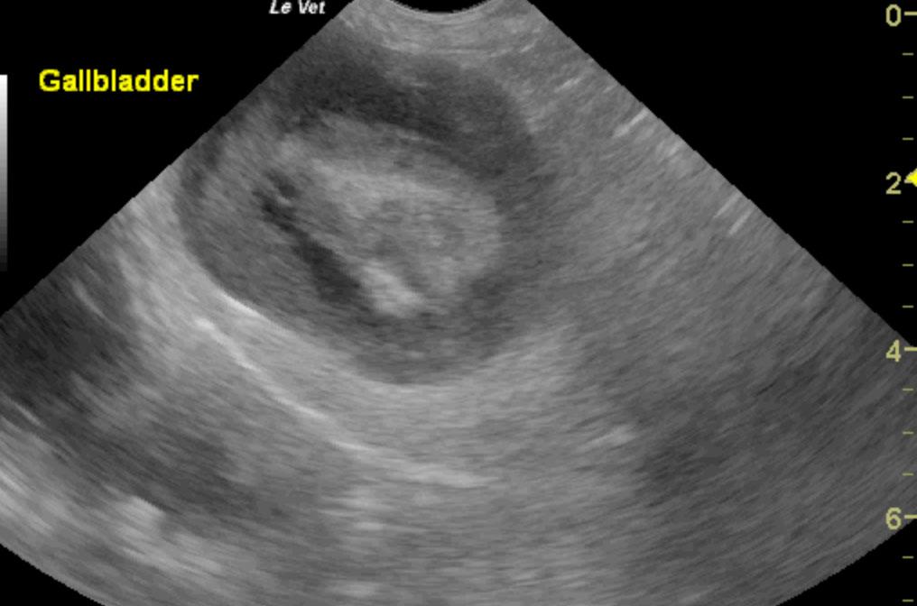
Increased echogenic appearance of the liver. Full gall bladder containing hyperechoic sludge-like material. Bile duct not visualized.
DX
Outcome
The patient was euthanized on the table during surgery. There were already a lot of adhesions around the liver and gallbladder, and the very distended gallbladder ruptured spectacularly right at the junction of the cystic duct while trying to free the adhesions. The cystic duct was also completely occluded.
Clinical Differential Diagnosis
Liver: neoplasia, abscess, granuloma, bacterial/fungal hepatitis, toxins, nodular hyperplasia Gall bladder: cholecystitis, mucocele, rupture, neoplasia, obstruction (sludge, lith, duodenal/pancreatic disease)
Sampling
None
Video
Patient Information
Clinical Signs
- Anorexia
- Diarrhea
- Lethargy
- Vomiting
Images
Blood Chemistry
- Albumin, Low
- Elevated Liver Enzymes
- Glucose, Low
- Total Bilirubin, High
CBC
- Basophils, High
- Left Shift
- Neutrophils, High
Clinical Signs
- Anorexia
- Diarrhea
- Lethargy
- Vomiting