An 8 year old NM Maltese was presented for acute onset anorexia and being depressed. The only significant abnormality on urinalysis, CBC, and serum biochemistry was severely elevated ALT and ALP activity.
An 8 year old NM Maltese was presented for acute onset anorexia and being depressed. The only significant abnormality on urinalysis, CBC, and serum biochemistry was severely elevated ALT and ALP activity.
Case Study
03_00223 Cody P Mucocele
Sonographic Differential Diagnosis
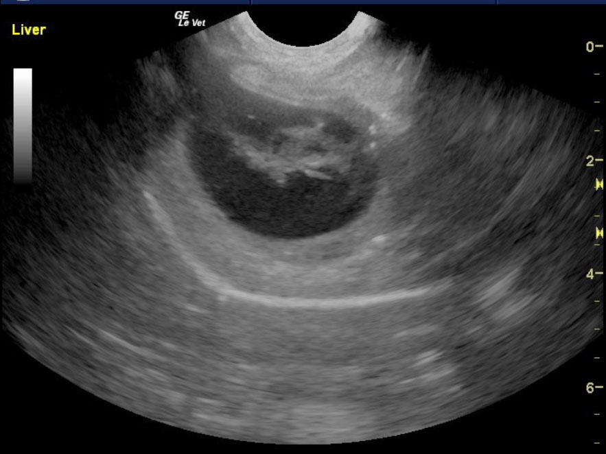
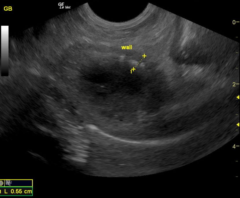
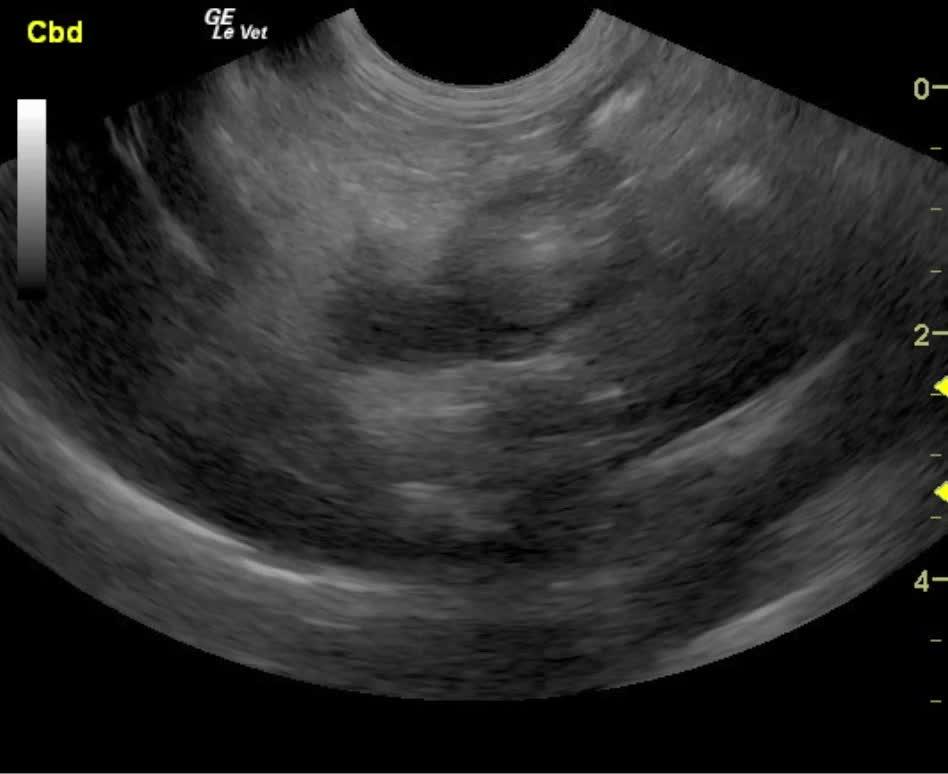
Inflamed and adhered gallbladder mucocele with adhesion pattern in the region of the porta hepatis. Minor common bile duct dilation. Recommend cholecystectomy in this patient, common bile duct lavage, hepatic and gastrointestinal biopsies would be ideal. I do not suggest medical treatment in this patient given that there are no signs of gallbladder viability given the suspended, immobile debris. No material within the gallbladder appeared to be moving. Therefore, this gallbladder is essentially dysfunctional. In addition the adhesion pattern suggests history of perforation and reaction from the body to wall. Therefore, cholecystectomy and common bile duct lavage is the only long term solution along with liver biopsy. Cholangiohepatitis protocol would prove fruitful in the short-term. However, long term resolution would require surgical intervention.
Image Interpretation
The gallbladder in this patient presented double layered wall with focal mineralization or potential gas penetration of the wall with aggressive adhesion pattern at the neck. Dilated gallbladder neck and suspended immobile debris were also noted. This is consistent with gallbladder mucocele and secondary inflammation. The patient had a positive Murphy’s sign which was reported to the clinician by the sonographer. No free fluid was noted at this time. The common bile duct did appear slightly dilated and occasional echogenic structure was noted within the common bile duct, which may result in biliary calculi. The gallbladder measured 4 x 3 cm and was significantly inflamed. Surgical intervention with cholecystectomy and common bile duct lavage would be warranted. There is a minor possibility that bile duct deviation would be required. The common bile duct proximal to the duodenal papilla measured 0.32 cm. However, an adhesion pattern was noted in this region. The gallbladder wall measured 0.55 cm. The liver itself presented increased portal markings with heterogenous changes. This is consistent with cholangiohepatitis pattern.
DX
Mucocele
Outcome
Cholecystectomy was performed.
Clinical Differential Diagnosis
Acute liver disease – toxins, infectious (bacterial/viral/fungal), trauma, secondary to pancreatitis/peritonitis/gall bladder disease Gall bladder – obstruction, mucocoele, cholecystitis, rupture
Sampling
Culture of the gallbladder was performed and no aerobic growth was noted. The liver and gallbladder both did not reveal any evidence of neoplasia.
Video
Patient Information
Gender :
Male, Neutered
Species :
Canine
Type of Imaging : Ultrasound
Status :
Complete
Clinical Signs
- Anorexia
- Lethargy
Images
Blood Chemistry
- Alkaline Phosphatase (SAP), High
- ALT (SGPT), High
Clinical Signs
- Anorexia
- Lethargy