An 8-year-old MN cat presented for anorexia and lethargy. Physical examination revealed minor dehydration but otherwise was normal. CBC and blood chemistry profile revealed severe ALT and AST elevations with mild regenerative anemia, rouleaux formation, and moderate neutrophilic leukocytosis with a left shift. Urinalysis was unrevealing.
An 8-year-old MN cat presented for anorexia and lethargy. Physical examination revealed minor dehydration but otherwise was normal. CBC and blood chemistry profile revealed severe ALT and AST elevations with mild regenerative anemia, rouleaux formation, and moderate neutrophilic leukocytosis with a left shift. Urinalysis was unrevealing.
Case Study
03-00093 Buster N Hepatic adenoma–RESEARCH ONLY–
Sonographic Differential Diagnosis
A potentially aggressive neoplasm partially impeding hepatic venous return was considered primarily. Inflammatory disease was considered unlikely.
Image Interpretation
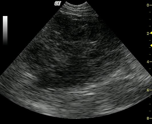
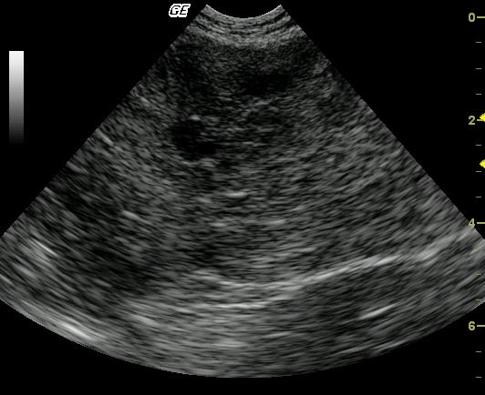
Long-axis view of the left lateral liver lobe reveals a large (5-cm), lobulated, complex, mixed echogenic, nodular mass arising from the left caudal liver. The mass is causing caudal displacement of the stomach, which was not in the view. The remaining left liver demonstrates a mildly echogenic parenchyma with evidence of portal venous congestion.
DX
Underlying hepatic adenoma with marked necrosis and inflammation
Outcome
The patient thrived after surgery, with all hepatic enzymes returning to normal after 2 months of outpatient care. This patient was clinically normal 6 months after surgery.
Comments
No video is available on this patient.
Clinical Differential Diagnosis
Inflammatory hepatopathy, pancreatitis, IBD, biliary calculi, neoplasia, tricobezoar or other GI obstruction.
Sampling
18-gauge US-guided biopsy revealed acute focal hepatic necrosis and infarction. Surgical resection revealed liver lobe torsion, whereas surgical biopsy evidenced an underlying hepatic adenoma with marked necrosis and inflammation. Thoracic radiographs were free of lesions consistent with metastatic disease.
Patient Information
Gender :
Male, Neutered
Species :
Feline
Type of Imaging : Ultrasound
Clinical Signs
- Anorexia
- Lethargy
Exam Finding
- Dehydration
Images


Blood Chemistry
- ALT (SGPT), High
- AST (SGOT), High
CBC
- Left Shift
- Neutrophils, High
- RBC, Low
- WBC, High
Clinical Signs
- Anorexia
- Lethargy