– 9 yr old FS Portugese Water dog decreased appetite and vomiting for about 1 week
– mild elevation in ALT, neutrophilia, cPLI SNAP negative; abdominal rads unremarkable
– liver normal on u/s (have recommended pre/post bile acids)
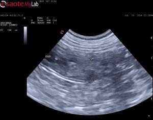
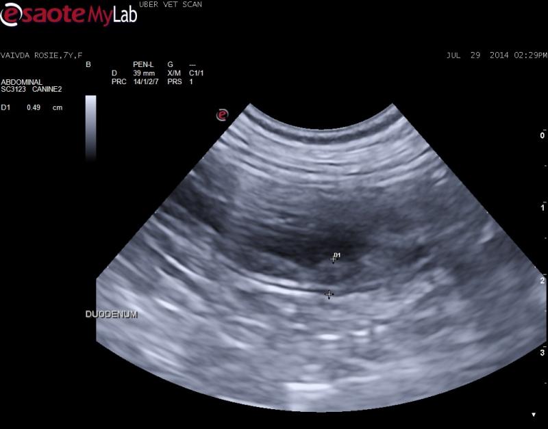
– mucosal layer of duodenum and upper GI appears thick and hyperechoic – possible luminal mass with loss of normal layering
– no lympadenopathy and distal GI, colon appears normal
What do you think about this bowel?


Comments
Nice image set Jacquie,
Nice image set Jacquie, for most of the bowel the lesions are mucosal but for about 2 cm the lesion goes transmural but the serosa is minimally affected. I would cut it out as carcinoma does this and the key tis to get carcinoma out early. However intraoperative ultrasound is key here to 1) find the lesion since no serosal changes for the surgeon to see easily because at the moment th epathology is mural and luminal and 2) to delineate solid healthy layered intestine to R&A. Other things that do this are focal ibd/granulomatous, spontaneous necrosis and focal complicated enteritis… maybe lsa but doubt it. Also be sure its there right before sx so it doesn’t “heal itself” before your go in.
More on intraoperative US here:
http://sonopath.com/resources/interventional-procedures