This 7 year old F Beagle presented initially after episode of collapse. Recurrent thoracic effusion. Mildly elevated WBC, all else wnl
Case Study
CT – Recurrent pyothorax with lymphadenitis in a 7 year old F Beagle
Image Interpretation
CT of the thorax –
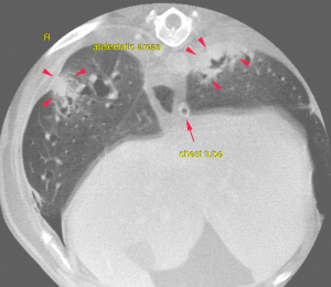
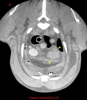
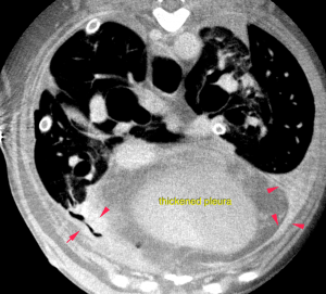
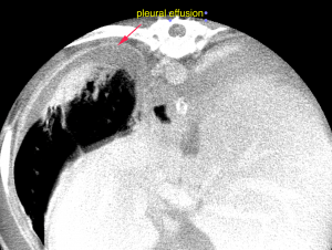
Both pleural cavities present a mild amount of non-contrast enhancing gravity dependent uniformly soft tissue attenuating fluid. The generalized retraction and cortication of the lung and pleural thickening has progressed. The lung presents with multiple surface retractions associated with pleura-based interstitial bands emerging from the visceral pleura (pleural and interstitial scarring). The pleura is markedly thickened and presents mildly irregular with multifocal small mineralized foci. The sternal and cranial mediastinal lymph nodes are moderately enlarged with maintained short to long axis ratio of < 0.5.
DX
The findings are consistent with recurrent pyothorax
Outcome
Mild bilaterally symmetric pleural effusion with marked pleuritis and progressing pneumonitis scarring • Secondary reactive/suppurative mediastinal lymphadenitis • Pulmonary osteomas & pleural plugs No evidence of a migrating foreign body was identified. As no structural cause is identified a past traumatic puncture injury with bacterial seeding is likely to be the underlying pathomechanism. Consider repeated warm saline lavages and systemic broad spectrum antimicrobial treatment as initial conservative treatment option. If clinical signs persist a surgical option is warranted either by thoracotomy or by thoracoscopy.
Patient Information
Gender :
Female, Intact
Species :
Canine
Type of Imaging : CT
Clinical Signs
- Collapse
Images
CBC
- WBC, High
Clinical Signs
- Collapse