Left adrenal mass invasion, likely pheochromocytoma, in a 10 year old MN Labrador Retriever

Case Study

Left adrenal mass invasion, likely pheochromocytoma, in a 10 year old MN Labrador Retriever

A 10-year-old NM Labrador cross was presented for inappetence, abdominal pain, heavy breathing, and intermittent abdominal distension. Blood work was normal. Survey radiographs showed splenomegaly and decreased liver size. 
A 10-year-old NM Labrador cross was presented for inappetence, abdominal pain, heavy breathing, and intermittent abdominal distension. Blood work was normal. Survey radiographs showed splenomegaly and decreased liver size. 
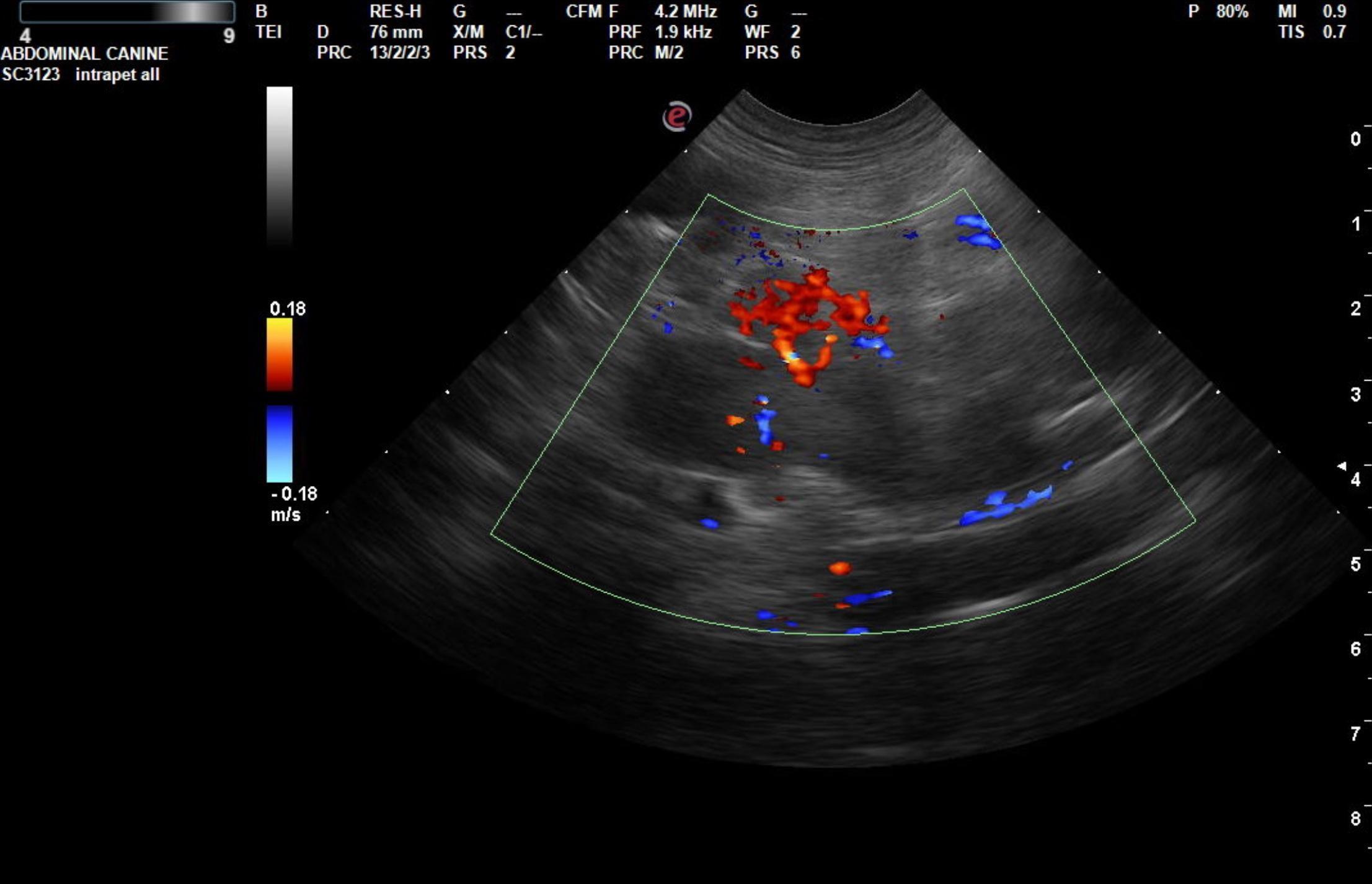
Image Interpretation

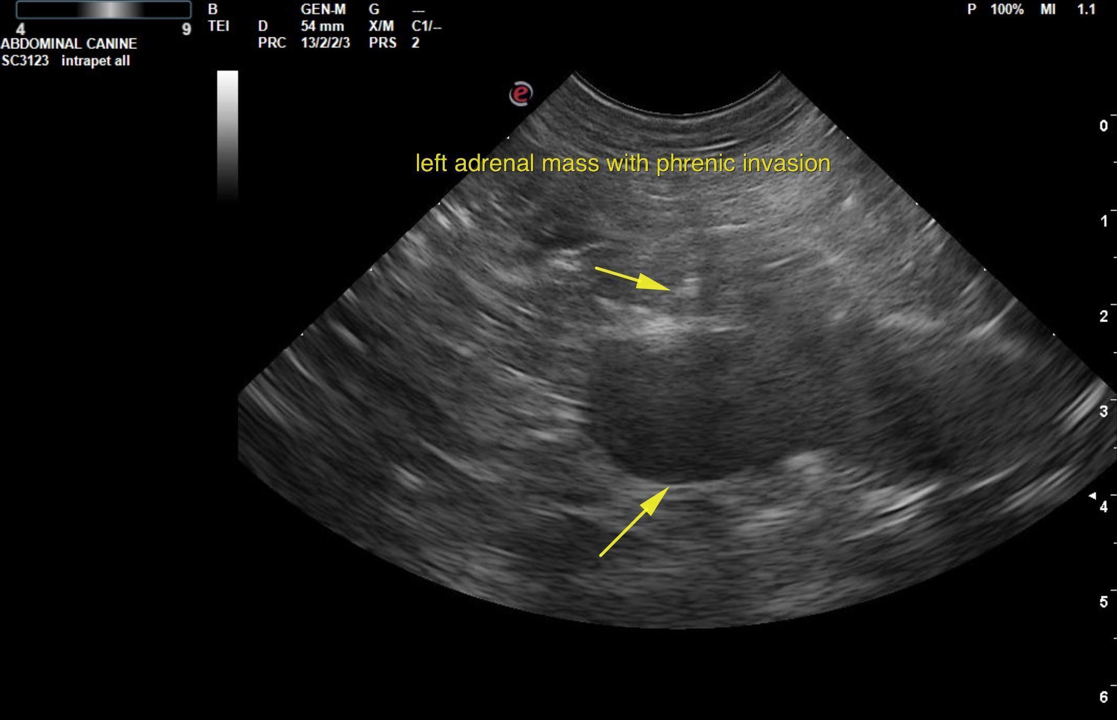
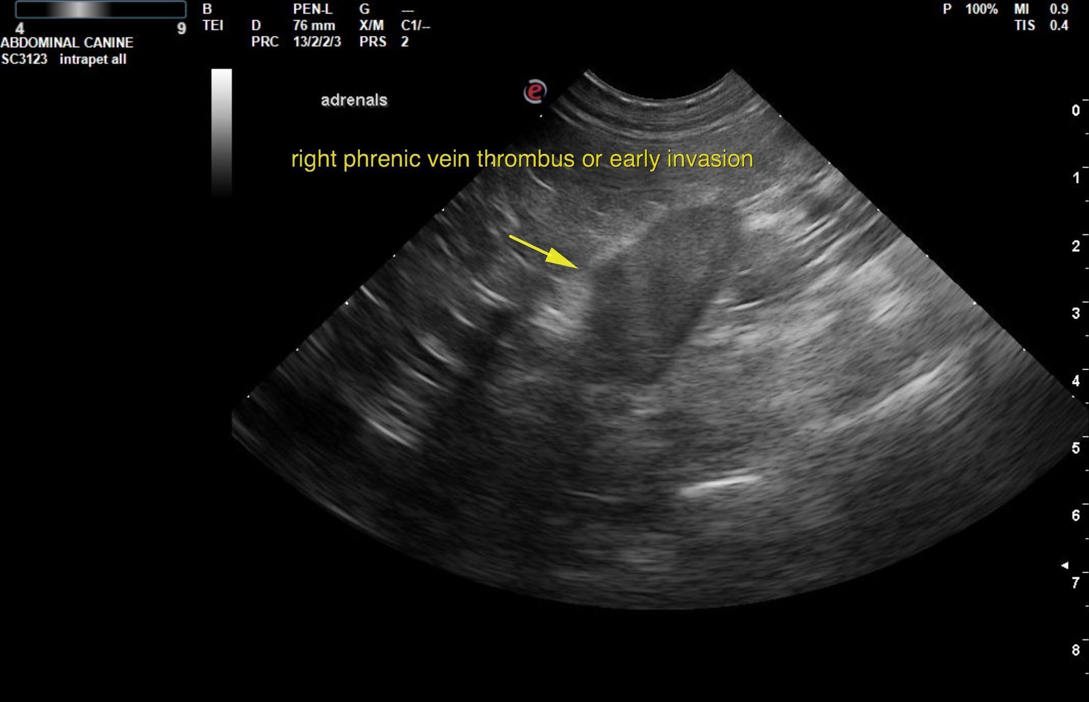
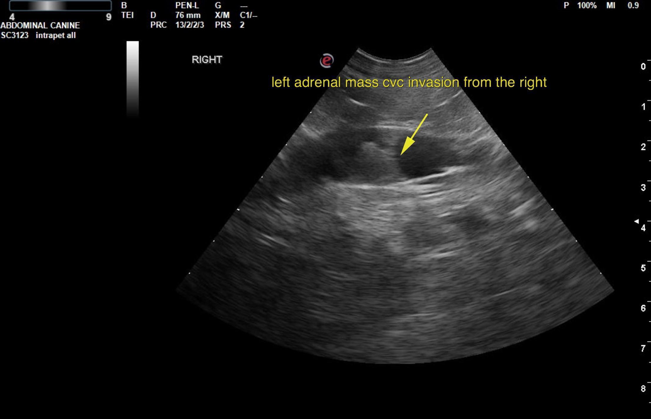
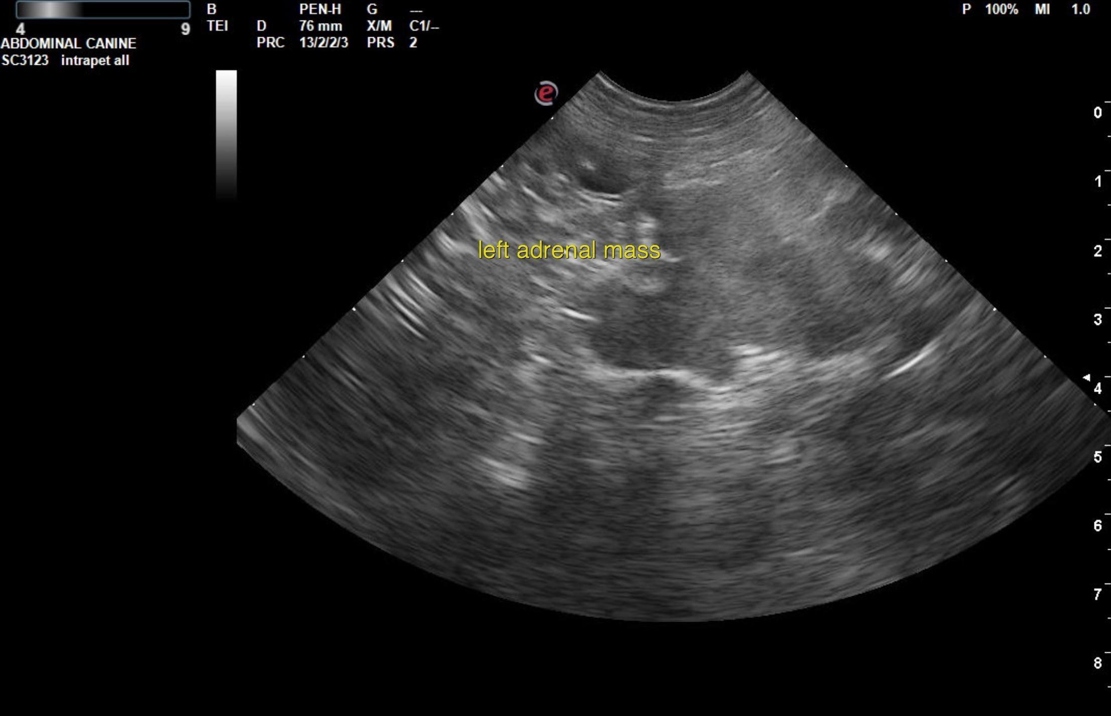
The left adrenal gland presented an irregular mass with phrenic vein invasion. The mass measured approximately 4.0 cm. The mass was significantly vascular. The left adrenal mass invaded the phrenic vein for approximately 2.0 cm.  The right adrenal gland was uniformly enlarged and measured 3.0 x 2.0 cm with loss of corticomedullary definition.

The liver images from right and left intercostal as well as subcostal views revealed subjectively normal liver size, contour, and structure. Some age-related parenchymal remodeling was noted but likely not clinically significant at this time. Vascular and biliary tracts were of normal volume and no evidence of congestion was noted. The gallbladder revealed a minor amount of excessive debris with an echogenic wall. 

DX

invasive left adrenal mass, Enlarged right adrenal gland. The phrenic vein appeared to slightly occupied on the right, potentially owing to phrenic vein thrombosis or early invasion from a dual neoplastic process. Age related changes elsewhere with chronic cholecystitis gallbladder pattern and excessive debris.

Outcome

Recommend serial blood pressure measurements in this patient. This is strongly consistent with pheochromocytoma with a potential for adenocarcinoma. Hyperplasia of the right adrenal gland is most likely. There was no evidence of invasion from the right. Surgical consultation is recommended.

Clinical Differential Diagnosis

Spleen – neoplasia, splenitis, torsion, hypersplenism
Abdomen – neoplasia, peritonitis, gastro-intestinal motility disorder

Patient Information

Patient Name : Cody Kruger/Bayshore
Gender : Male, Neutered
Species : Canine
Type of Imaging : Ultrasound
Status : Complete
Liz Wuz Here : Yes
Code : 07_00198

Clinical Signs

  • Abdominal Distension
  • Abdominal Pain
  • Anorexia
  • Dyspnea

Exam Finding

  • Splenomegaly

Images

krug3krug6kruig5krug10rb9

Clinical Signs

  • Abdominal Distension
  • Abdominal Pain
  • Anorexia
  • Dyspnea