Ultrasound of the left and right achilles –
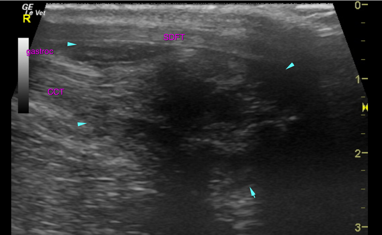
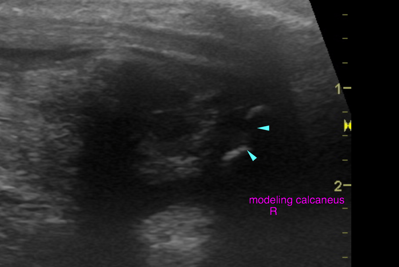
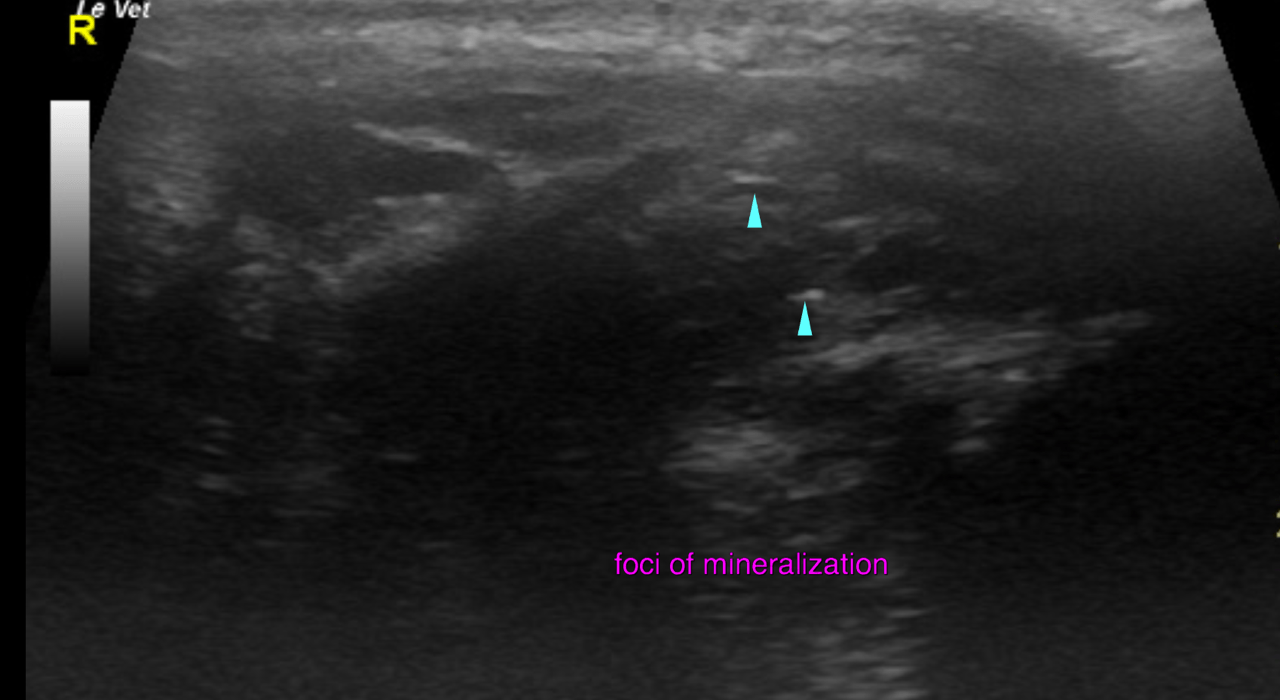
Right:
Both portions of the deep calcaneal tendon – the gastrocnemius and the common calcaneal tendon – present a severe focal swelling proximal to the calcaneus. The affected area extends 3 cm proximal from the calcaneus. The maximum diameter is 2.5 cm. Marked loss of the fibre pattern, decreased echogenicity and heterogeneity is noted. A mild amount of anechoic fluid is interspersed between the tendon layers. The calcaneus presents marked modeling with an irregular concave surface. Multifocal mineralization is seen within the gastrocnemius and common calcaneal tendon.
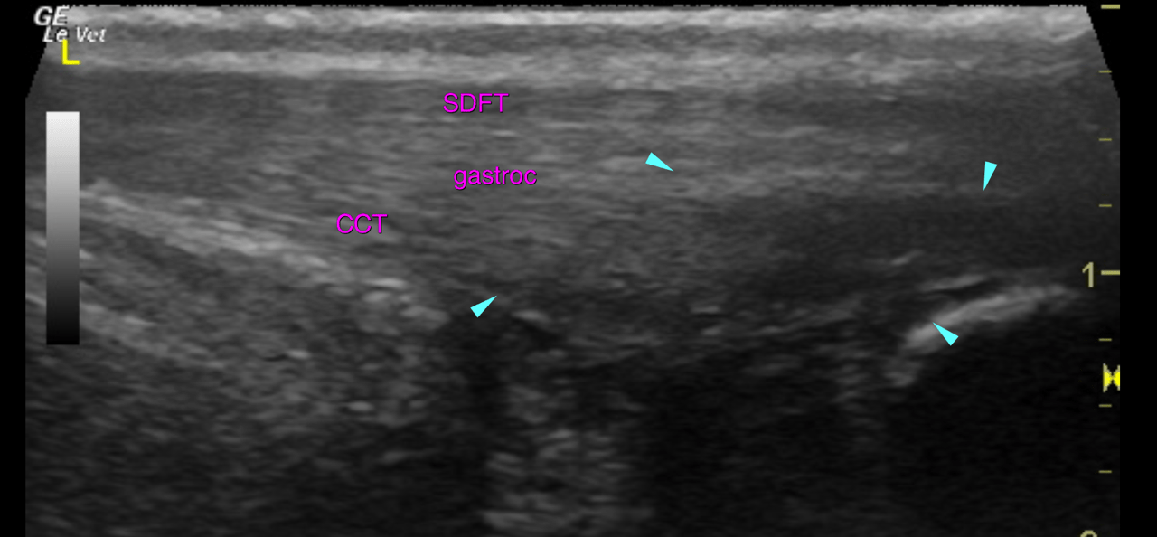
Left:
Similar but less advanced changes are noted for the left deep calcaneal tendon. The affected area extands 2 cm proximal to the calcaneus and is 1 cm in diameter. The loss of fibre pattern is advanced here too. However mineralization and bony modeling of the calcaneus are not seen here yet