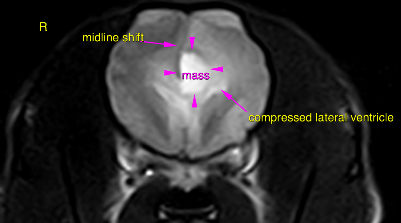
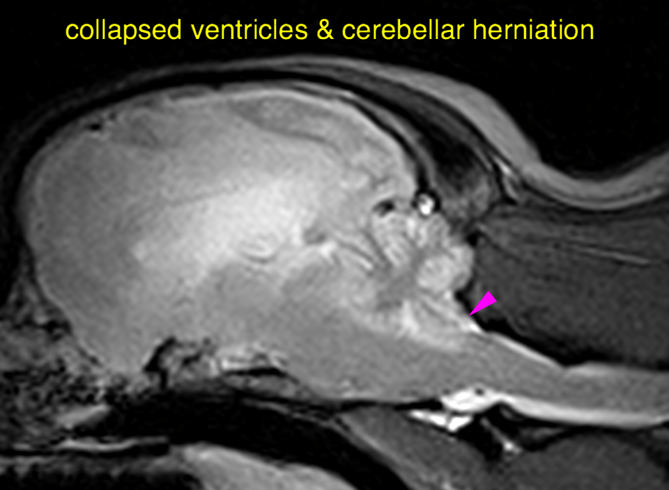
This 10 year old MN Boston Terrier dog has been ADR for the past few months, withdrawn, sedate, with developing hypermetric movements and loss of balance
Physical Exam: Obtunded; episodes of unawareness, ataxia and collapse. Murmur 1/6. Previous MRI found foramen magnum herniation of the cerebellum
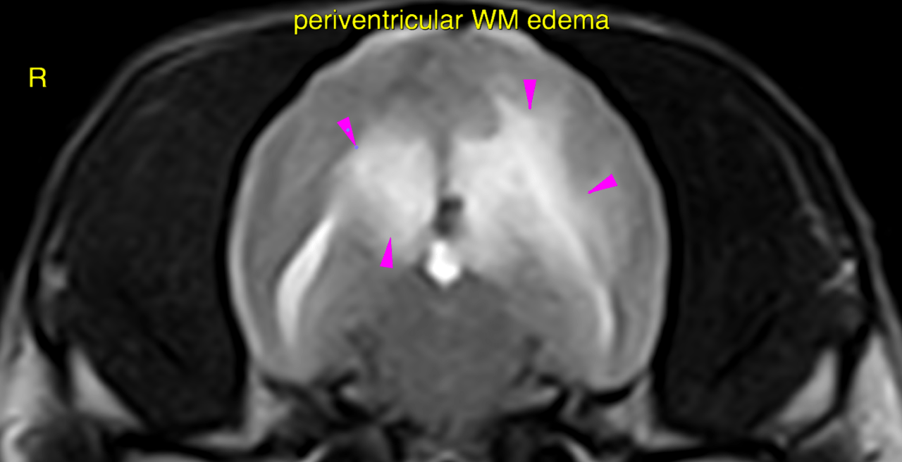
This 10 year old MN Boston Terrier dog has been ADR for the past few months, withdrawn, sedate, with developing hypermetric movements and loss of balance
Physical Exam: Obtunded; episodes of unawareness, ataxia and collapse. Murmur 1/6. Previous MRI found foramen magnum herniation of the cerebellum