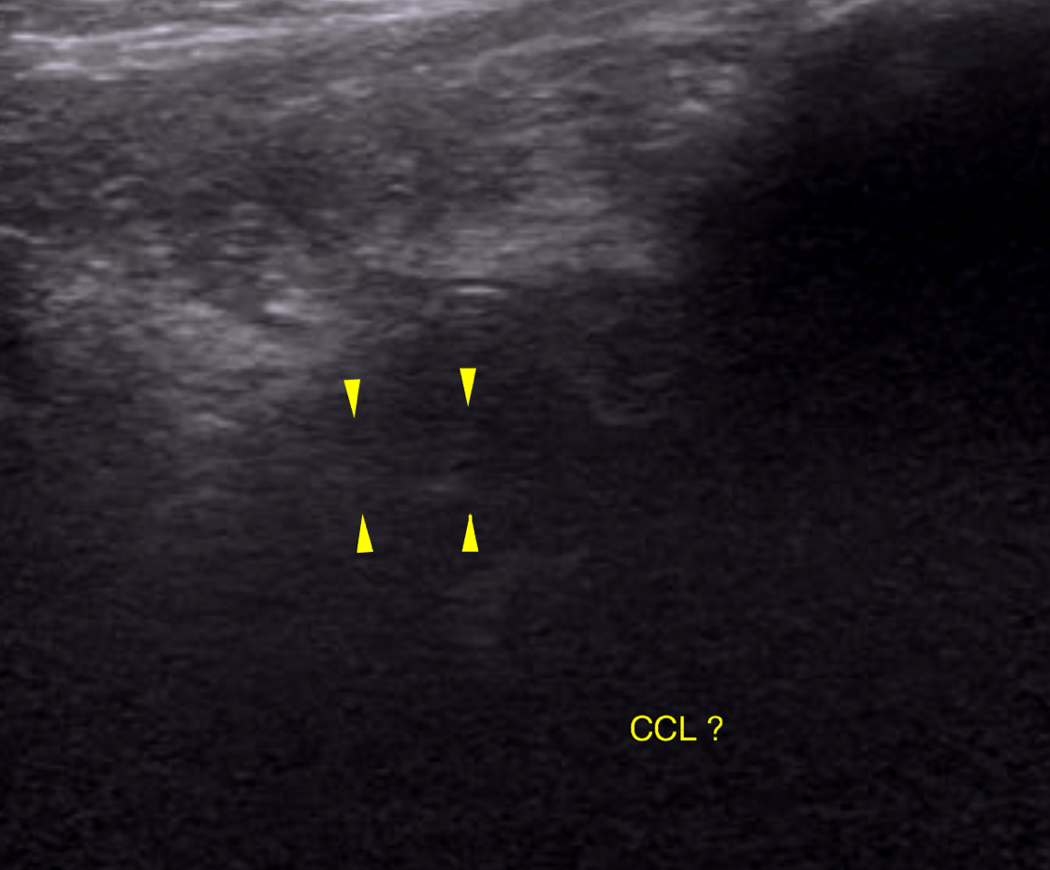
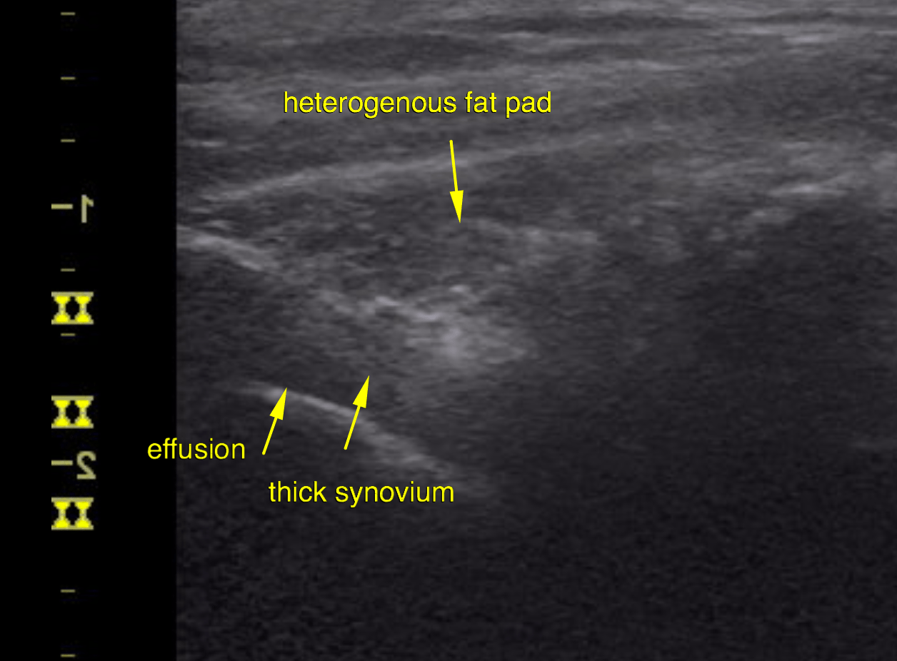
This FS Ridgeback mix dog has a history of bilateral medial luxating patella surgery. Was doing great then suddenly lame. Moderate cranial drawer sign left stifle
Physical Exam: WNL except for lameness LH leg
This FS Ridgeback mix dog has a history of bilateral medial luxating patella surgery. Was doing great then suddenly lame. Moderate cranial drawer sign left stifle
Physical Exam: WNL except for lameness LH leg