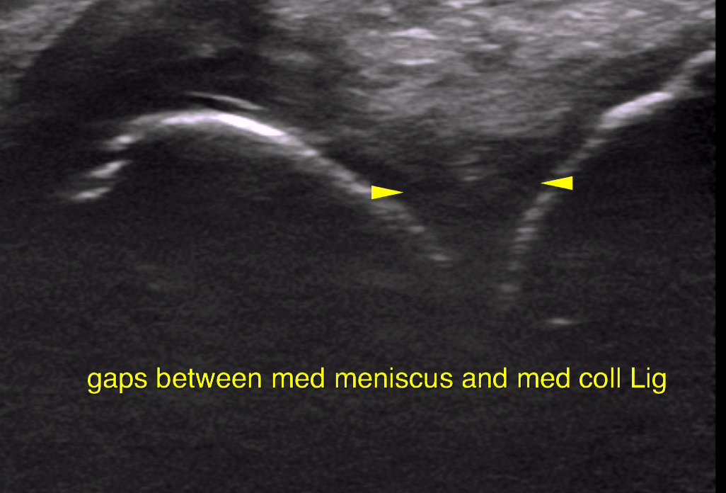
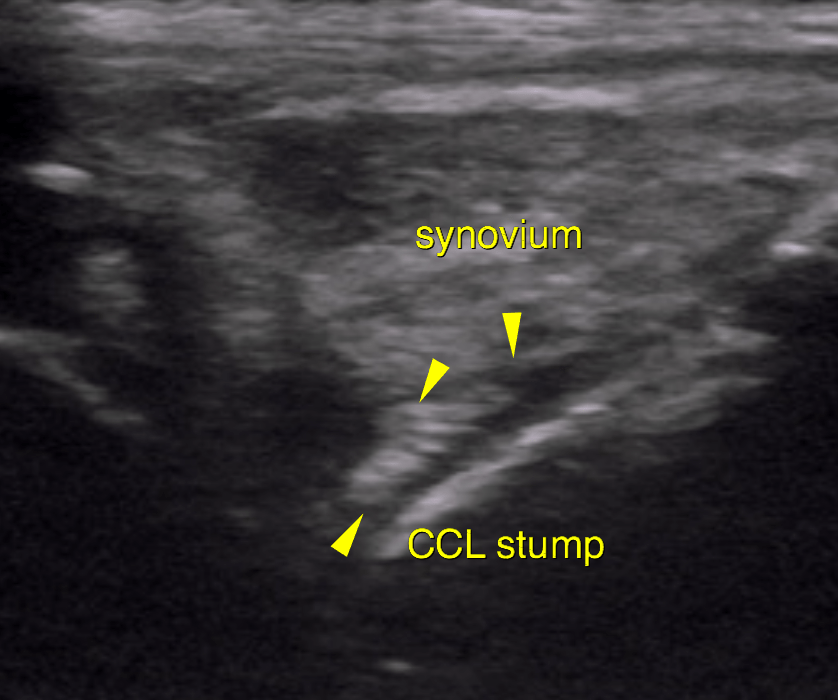
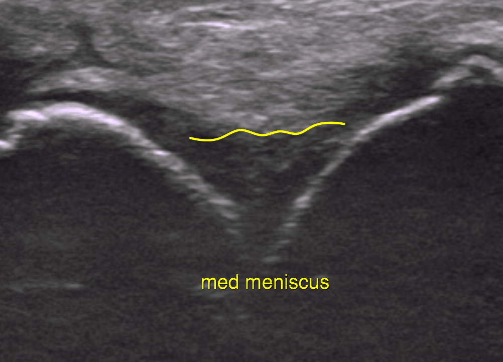
This 7 year old mixed dog presented with a history of lameness of the left rear leg on and off 1 month, stiff upon rising.
Physical Exam: Positive Cr drawer, positive CTT, pain left stifle, med buttress
CBC/Chem Pending.
Reason for Ultrasound Exam: Pre surgical scan
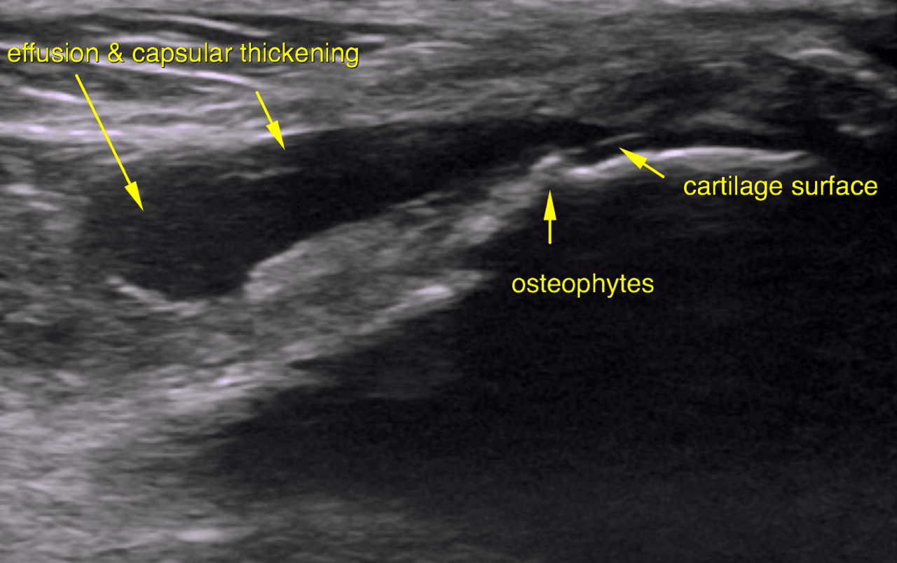
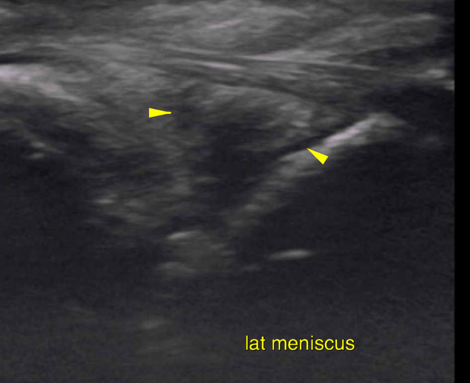
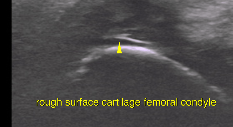
This 7 year old mixed dog presented with a history of lameness of the left rear leg on and off 1 month, stiff upon rising.
Physical Exam: Positive Cr drawer, positive CTT, pain left stifle, med buttress
CBC/Chem Pending.
Reason for Ultrasound Exam: Pre surgical scan