An 8-year-old FS DLH was presented for evaluation of vomiting and lethargy over a 2 days period. Renomegaly was present on abdominal palpation. Survey radiographs showed bilateral renoliths.
An 8-year-old FS DLH was presented for evaluation of vomiting and lethargy over a 2 days period. Renomegaly was present on abdominal palpation. Survey radiographs showed bilateral renoliths.
Moderate to severe hydronephrosis right kidney with corticomedullary calculi and probable right ureteral stricture. Obstructed left proximal ureter and mild to moderate hydronephrosis of the left kidney as well as cortical infarct and degenerative changes, further non-obstructive calculi were also noted. Minor bladder sand.
The urinary bladder presented small calculi with a minimal amount of shadowing. The pelvic urethra was imaged 2.0 cm beyond the cystourethral junction with no evident pathology noted. The right kidney in this patient presented severe hydronephrosis that measured 5.2 cm. Corticomedullary calculi were noted in the right kidney. The entrance to the right ureter in this patient was mildly dilated with echogenic debris. The cause of obstruction is not clear and this is likely owing to stricture. The distal right ureter was not dilated. The left kidney presented moderate swelling. A cortical infarct was noted at the cranial pole. Dilated renal pelvis and ill-defined pelvic fat were noted. Power Doppler assessment of the left kidney appeared to be hypervascular. The left ureter was obstructed approximately 1.0 cm distal from the left renal pelvis with a calculus.
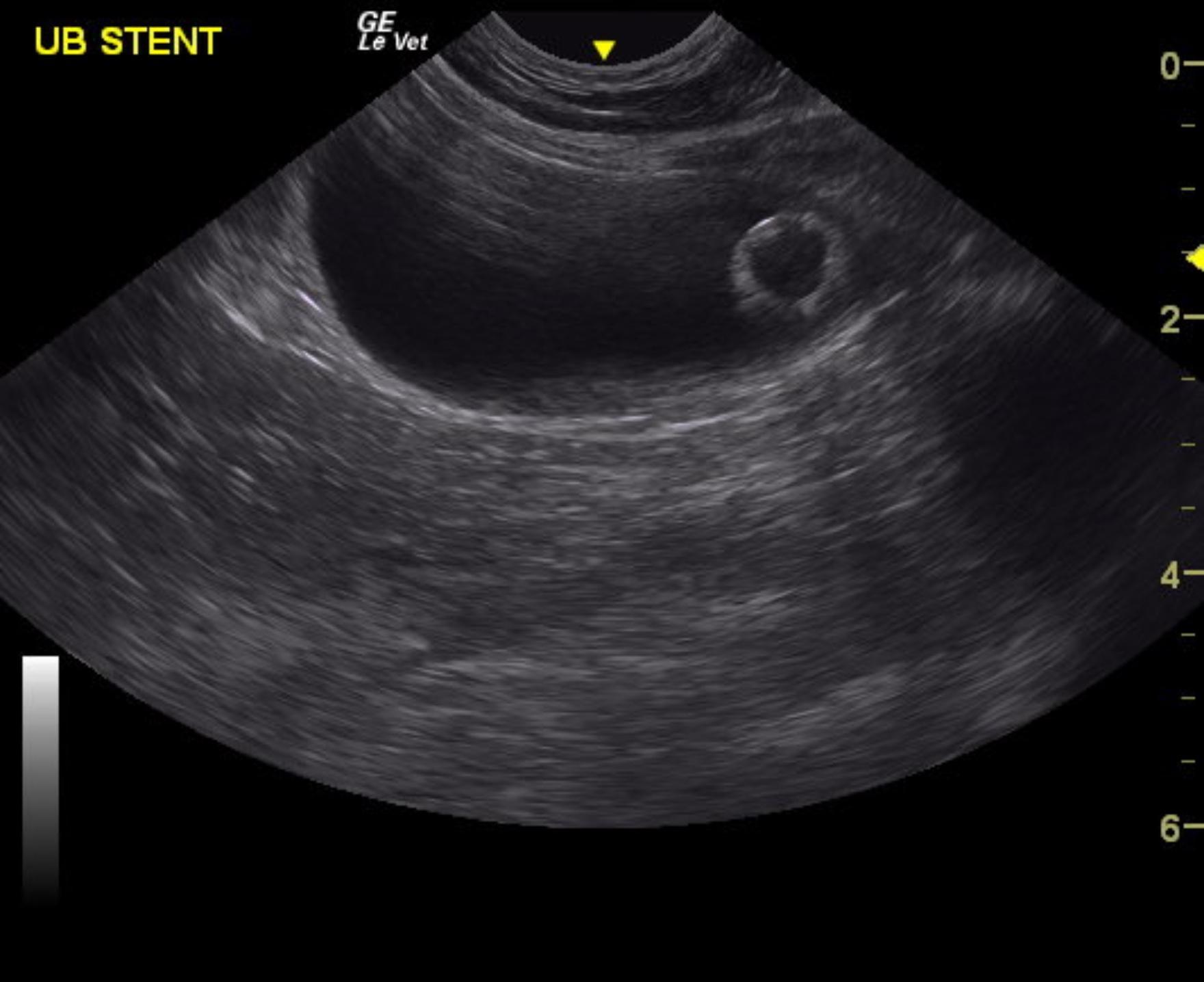
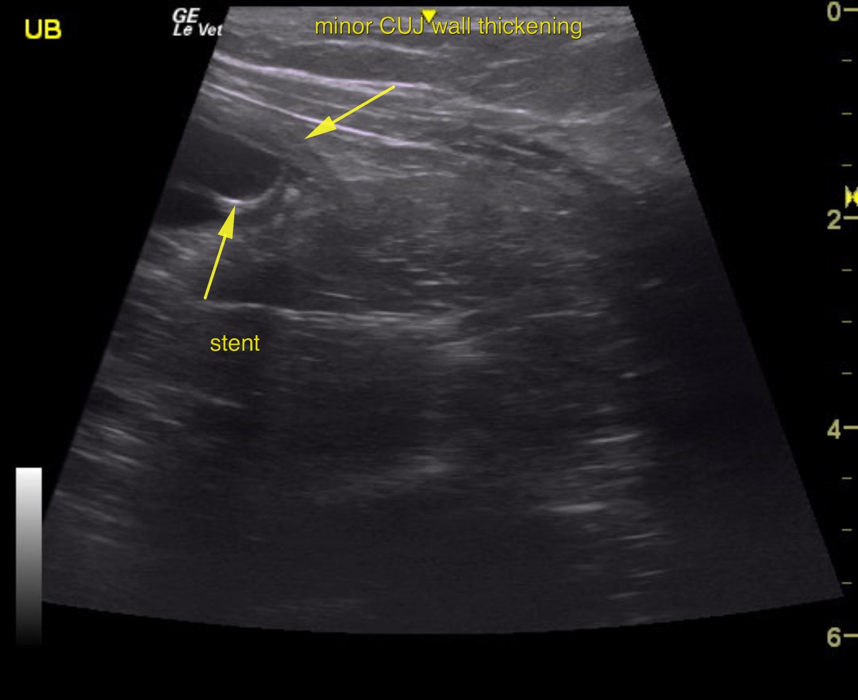
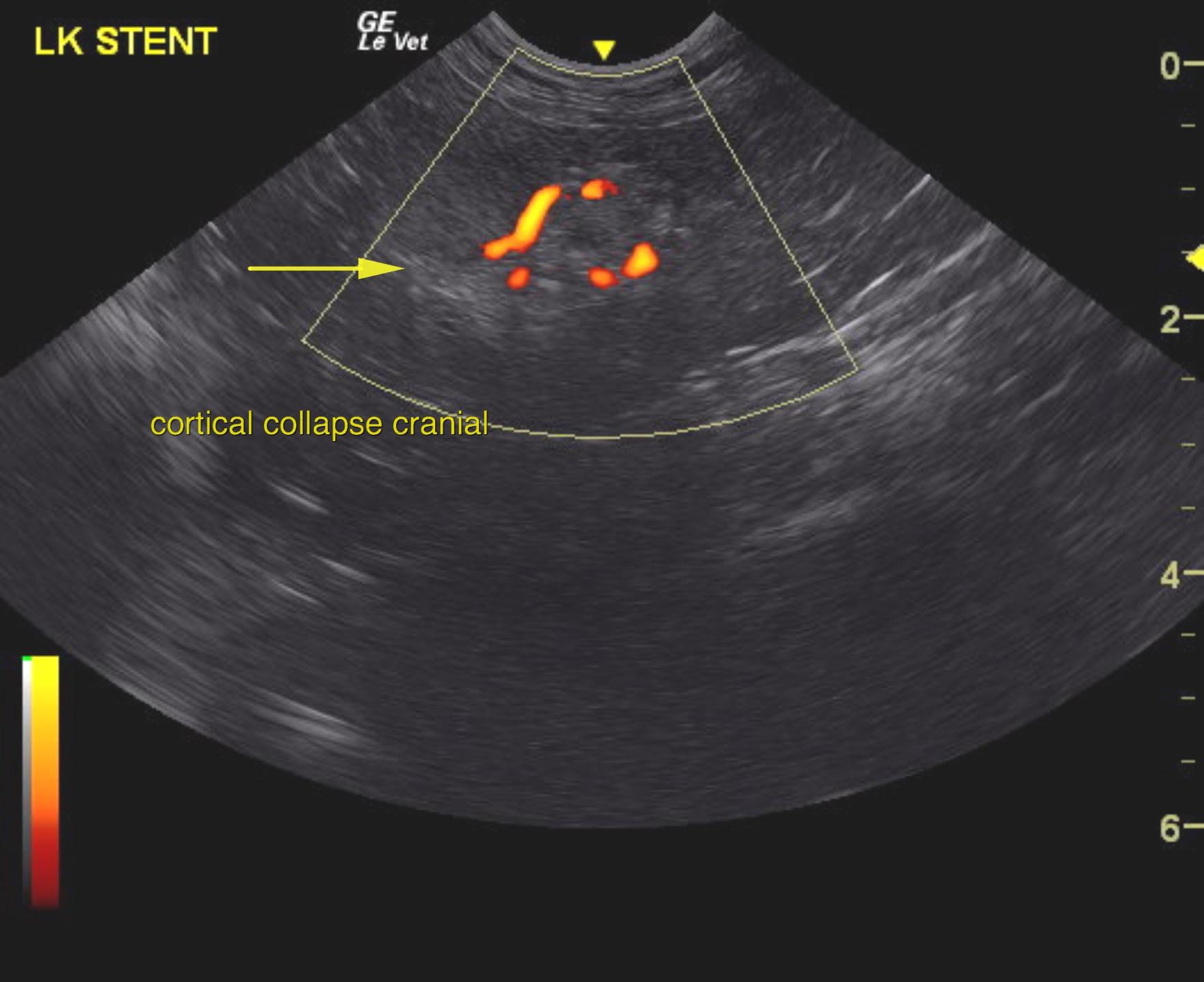
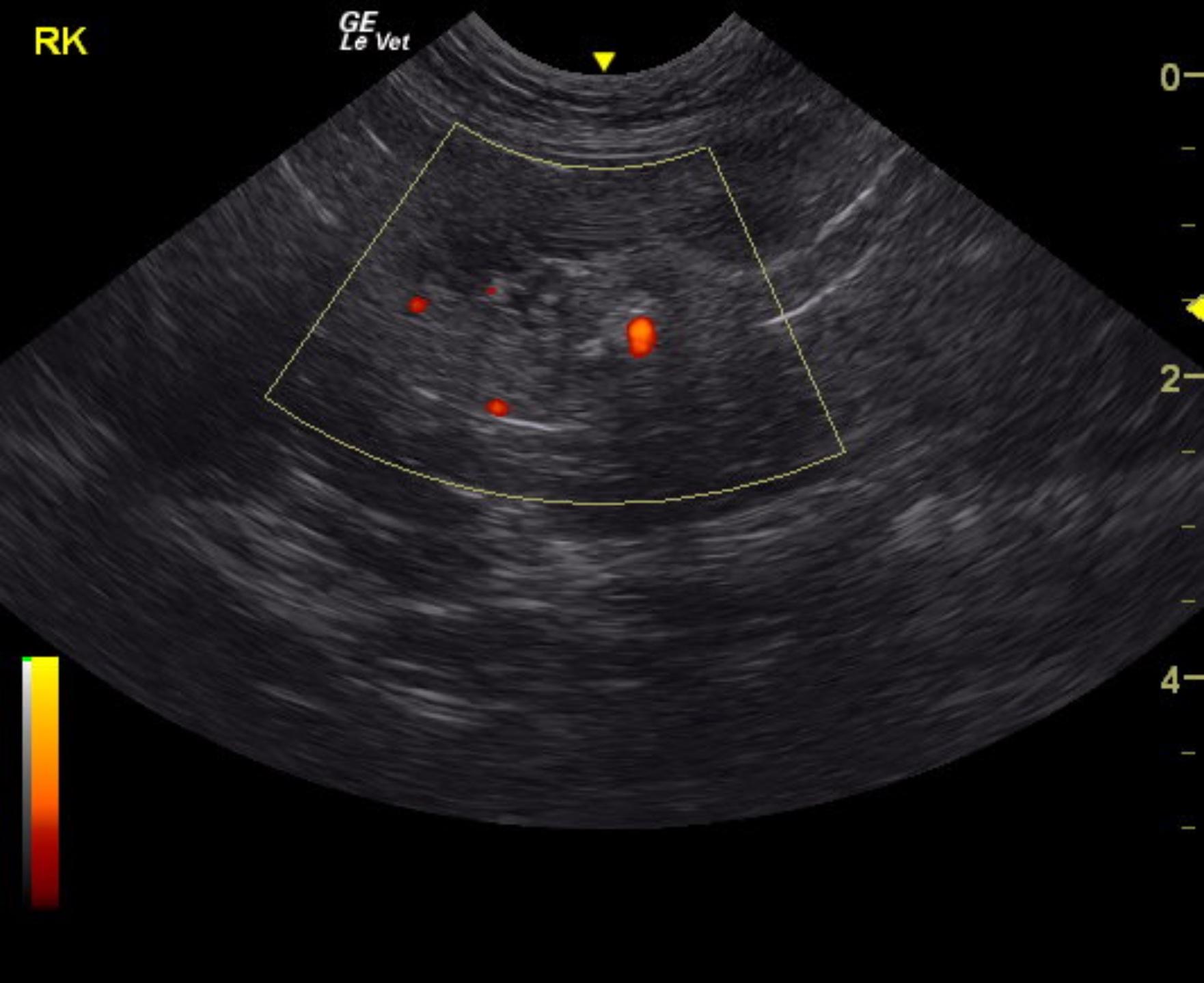
Three months later, the patient presented for check up on kidneys due to stent placement in the left ureter. The urinary bladder and ureters were unremarkable. However, there was slight thickening was noted at the cystourethral junction. This is likely hypertrophy secondary to stent placement or underlying mural disease. The left kidney in this patient presented moderate degenerative changes with interstitial nephrosis pattern, cortical remodeling and infarct, yet this is stable. The left kidney measured 3.51 cm. Power Doppler assessment of the left kidney appeared to be adequate to slightly subnormal. The right kidney was subnormal in size and measured 2.52 cm with cortical collapse owing to infarct. Minor corticomedullary mineralization was noted. Left kidney stent appeared to be in proper position with no evident inflammation. Diagnosis: Moderate degenerative renal changes with moderate to severe dystrophy of the left kidney. Cortical infarcts and collapse of both kidneys with slight pyelectasia of the left kidney.
Stent placement appears to be in proper position.
Pelvic calculi were noted in both kidneys and appeared to be stable.
Structurally stable patient.
Obstructive uropathy, renoliths, pyelonephritis, renal lymphoma, hydronephrosis
None