RAD – Mesenteric lymphadenopathy, lung lobe consolidation, splenomegaly and hypovolemia in a 6 year old FS Boxer dog

Case Study

RAD – Mesenteric lymphadenopathy, lung lobe consolidation, splenomegaly and hypovolemia in a 6 year old FS Boxer dog

This 6 year old FS Boxer dog presented with intermittent vomiting of one month duration. 

Physical Exam: icteric, increased liver values, vomiting, diarrhea, anorexia

CBC: normal

Chem: ALT 768, ALKP 593, GGT 38, Tbil 4.7, 4DX neg
This 6 year old FS Boxer dog presented with intermittent vomiting of one month duration. 

Physical Exam: icteric, increased liver values, vomiting, diarrhea, anorexia

CBC: normal

Chem: ALT 768, ALKP 593, GGT 38, Tbil 4.7, 4DX neg
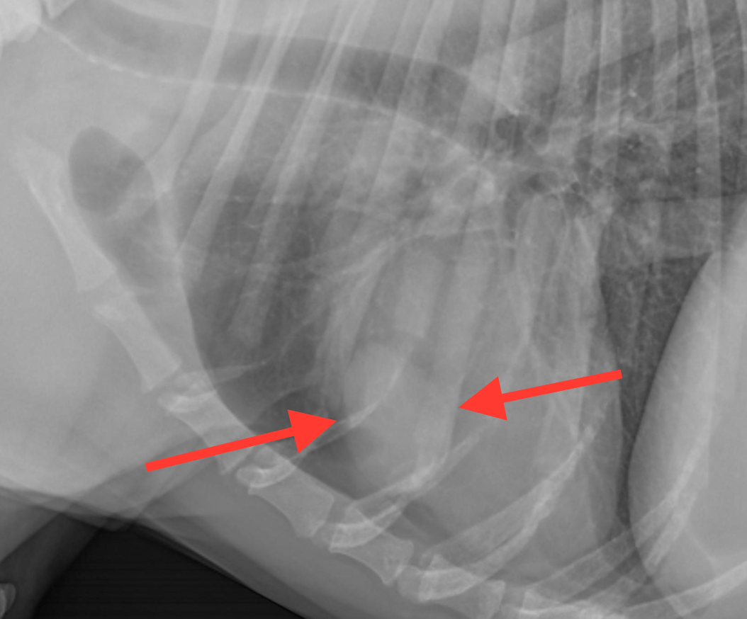
Image Interpretation

Rads of the thorax and abdomen – General:
Moderate degenerative changes of the axial skeleton are noted including spondylosis
and spondyloarthropathy.
Abdomen:
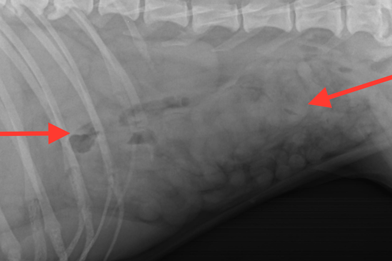
The serosal detail is significantly reduced in the mid and cranial abdomen. Crowding
of small intestinal loops with odd gas formations are noted in that area. The small
intestinal loops present nondilated and also even diameter throughout.
There is moderate generalized enlargement of the spleen with pointed margins.
The liver appears to have mildly rounded lobar margins but is normal for overall size.
The kidneys are within normal limits. The urinary bladder is mildly distended and
only partially seen cranial to the pubic rim.
Thorax:
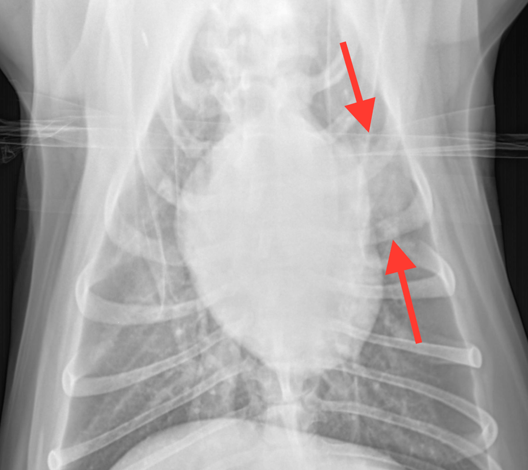
A regional peripheral alveolar lobar consolidation is noted for the cortical portion of
the left cranial lung lobe. There are no volume changes noted but the consolidation is
localized to a small portion of the lung lobe only.
There is hypovolemia with microcardia and underperfusion of the lung.

DX

Cranial and mid abdominal mesenteric lymphadenopathy/effusion. Peripheral and partial lobar consolidation of the left lung lobe. Splenomegaly. Hypovolemia.

Outcome

Disorders localized to the small intestine, mesenteric lymph nodes, pancreas, liver and
biliary system are possibly the underlying courses to the radiographic findings in the
abdomen.
Possible differential diagnoses for the lung changes include secondary neoplasia such
as round cell neoplasia (lymphoma, histiocytic sarcoma) and lobar pneumonia. With
the maintained lobar volume lobar atelectasis appears to be very unlikely. Because of
the involvement of multiple organs a multifocal round cell neoplastic infiltrate has to
be considered a potential in general here next to inflammatory disease.
To further define the underlying course of the radiographic changes abdominal
ultrasound and ultrasound of the lung changes with ultrasound-guided fine-needle
aspiration of the lung is strongly recommended.

Patient Information

Patient Name : Bella Reinstra/ACMP
Gender : Female, Spayed
Species : Canine
Type of Imaging : Ultrasound
Status : Complete
Liz Wuz Here : Yes

Clinical Signs

  • Anorexia
  • Vomiting

Exam Finding

  • Diarrhea
  • Icterus

Images

bildschirmfoto_2016-02-02_um_16bildschirmfoto_2016-02-02_um_16bildschirmfoto_2016-02-02_um_16

Blood Chemistry

  • Alkaline Phosphatase (SAP), High
  • ALT (SGPT), High
  • GGT High
  • Total Bilirubin, High

Clinical Signs

  • Anorexia
  • Vomiting

Special Testing

  • 4Dx Negative