06_00344 Buddy S Mineralizing prostatic and urethral mass

Case Study

06_00344 Buddy S Mineralizing prostatic and urethral mass

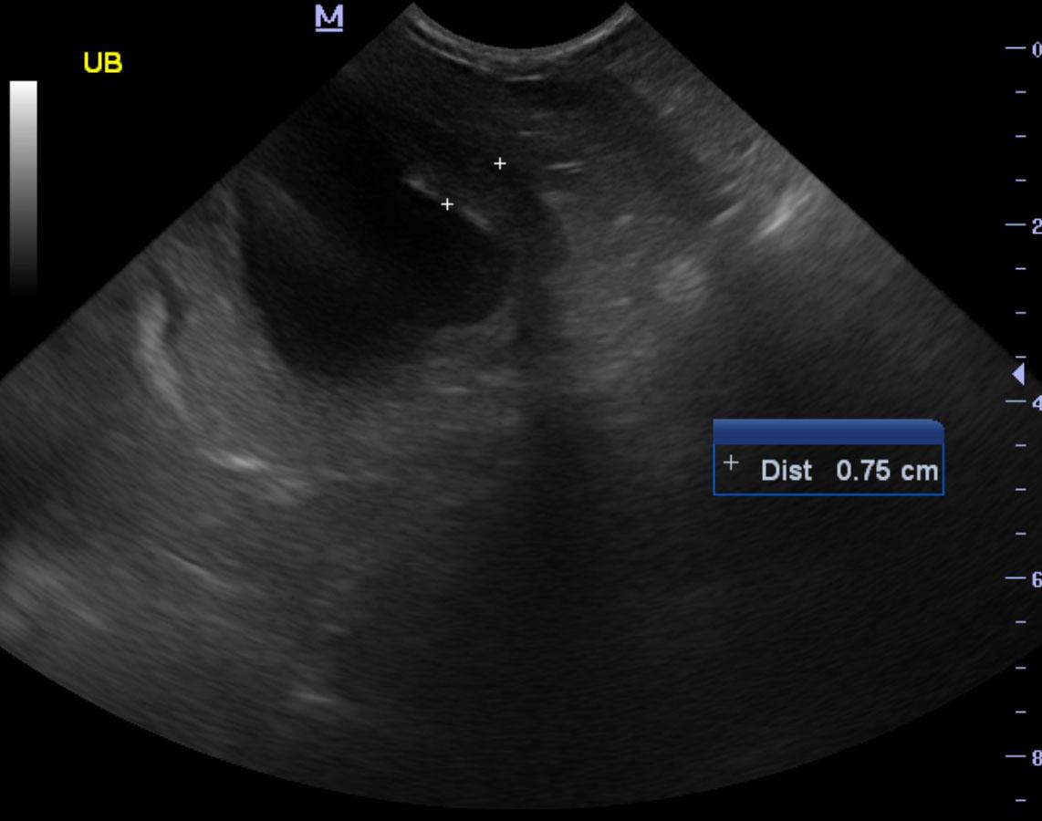
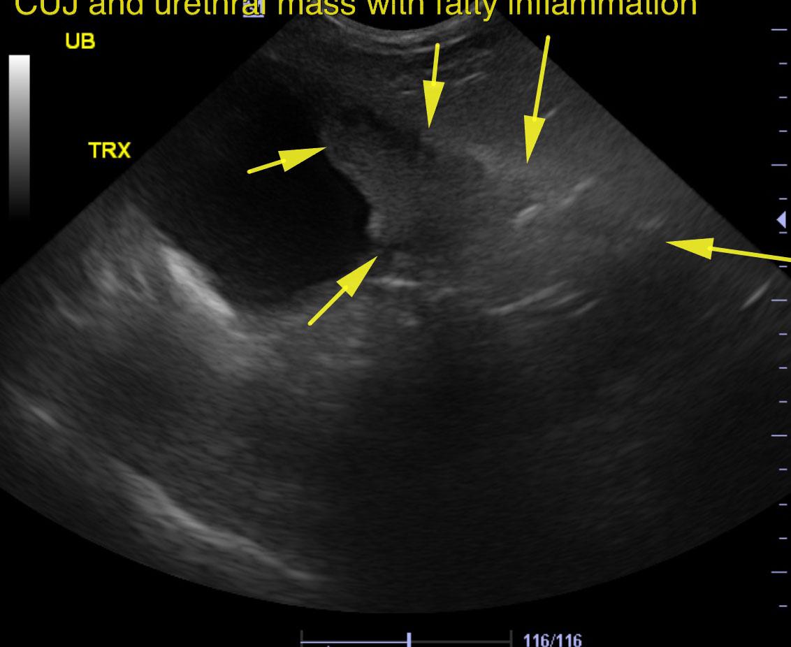
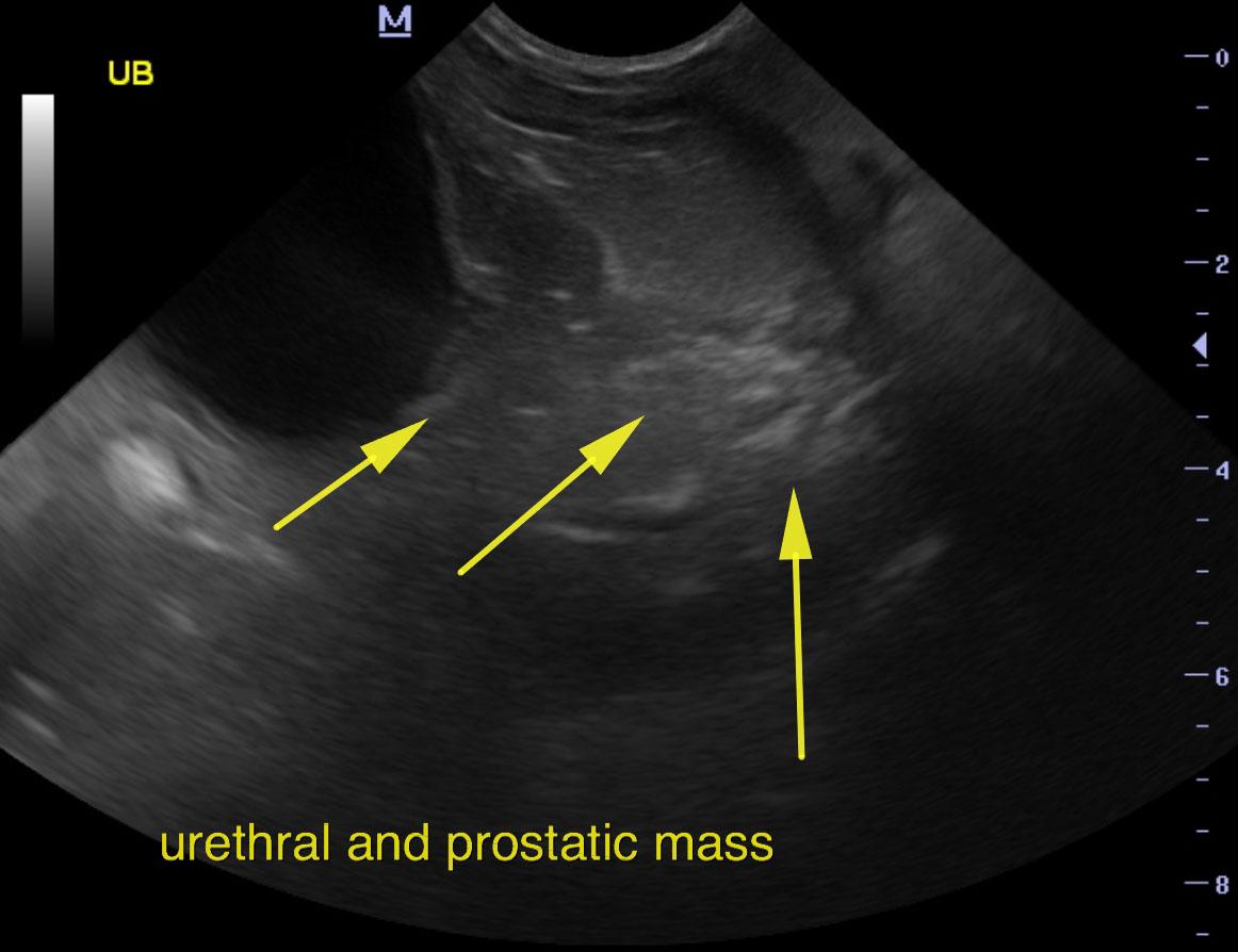
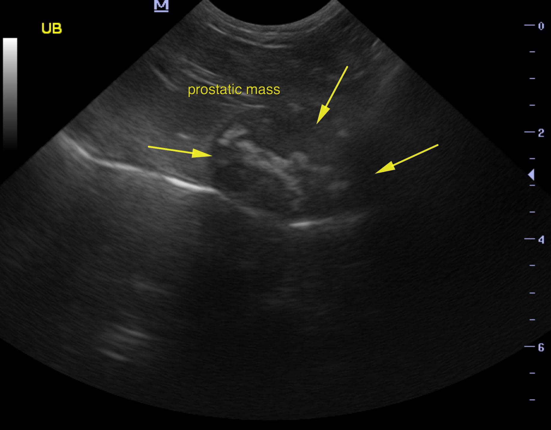
A 9-year-old NM Cocakpoo with a history of a calcium oxalate urolith being previously removed via cystotomy was presented for evaluation of recent on onset dribbling urine and stranguria that improved on antibiotic therapy. Physical examination was normal and a urinary catheter could be easily passed. Urinalysis showed a SG of 1.026 with trace of protein and negative culture.


Sonographic Differential Diagnosis

Mineralizing urethral mass that extended into the cystourethral junction and prostate. Concurrent bladder sand. Traumatic catheterization is recommended. Residual prostate appears to be involved. This is either prostatic or urethral in origin.

Image Interpretation

The urinary bladder presented a polypoid mass that occupied the pelvic urethra and cystourethral junction. The mass expands into the prostate, which measured approximately 2.0 cm with rounded, irregular contour and multi focal parenchymal mineralization. Periserosal inflammatory pattern was noted. Concurrent bladder calculi were also noted. Approximately 1.0 cm of sand accumulation was noted in the bladder, yet this is not the primary issue. The mineralizing urethral mass extends at least 3.0 cm into the urethra and expands approximately 1-1.5 cm into the pelvis with fatty inflammation.

DX

Mineralizing prostatic and urethral mass

Outcome

None

Clinical Differential Diagnosis

Bladder – urolith, neoplasia
Prostate – neoplasia, prostatitis

Sampling

None

UA Specific Gravity Range

1.026

Patient Information

Gender : Male, Neutered
Species : Canine
Type of Imaging : Ultrasound
Status : Complete

Clinical Signs

  • Incontinence
  • Stranguria

Images

jw3jw2jw5jw6jw4

Clinical Signs

  • Incontinence
  • Stranguria

Urinalysi

  • Culture negative
  • Protein Present