09_00164 Lunar W Prostatitis

Case Study

09_00164 Lunar W Prostatitis

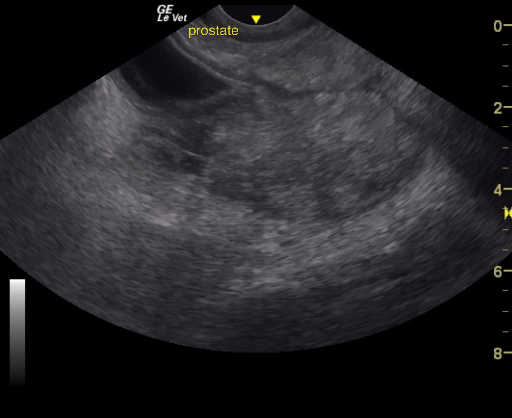
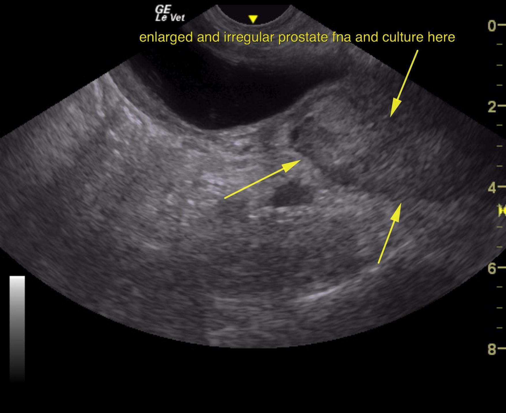
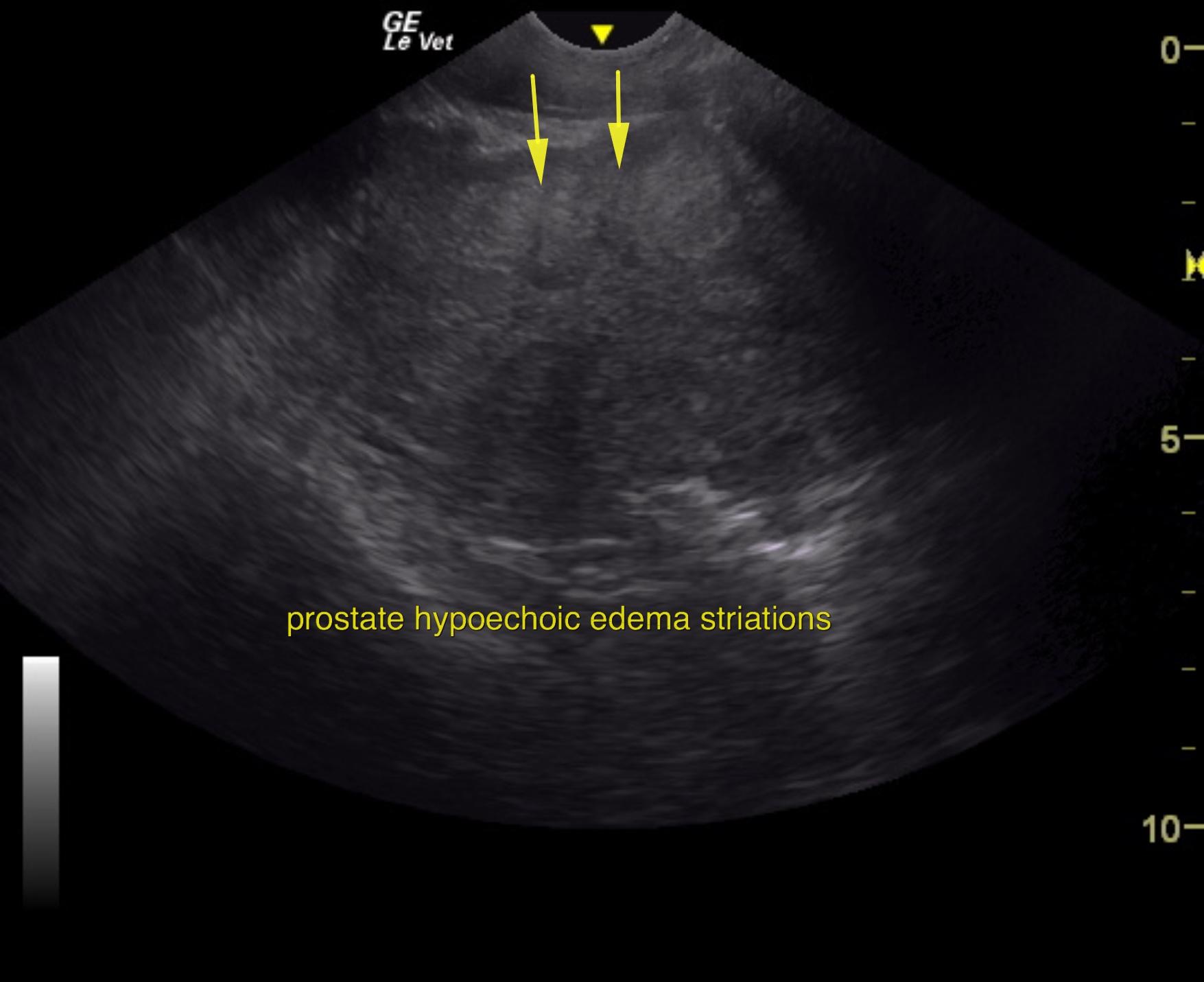
An 11-year-old NM Labrador with a history of gradual weight loss was presented for evaluation of abdominal pain and diarrhea. On abdominal palpation, the intestines were gas-filled, which was confirmed on survey radiographs. Elevated ALT activity, BUN, and glucose were present on serum biochemistry.


Sonographic Differential Diagnosis

Prostatitis, BPH presentation. There is a minor potential for neoplasia.

Image Interpretation

The prostate was enlarged and measured 5.0 cm.
A recheck exam was performed a week later. The prostate was persistently enlarged in this patient, nodular and irregular. Hypoechoic striations were noted within the prostatic parenchyma, suggestive for inflammation. The contour and nodular changes as well as the impingement upon the urethra has progressed compared to the prior sonogram with areas of localized fluid, potential microabscessation. Regional inflammation was noted. Deviation of the descending colon was noted in this patient and is likely the cause of straining.

DX

Prostatitis, BPH

Outcome

None

Clinical Differential Diagnosis

GIT – IBD, ulceration, neoplasia, dietary hypersensitivity, foreign body, intussusception
Pancreas – pancreatitis, neoplasia
Renolith
Peritonitis – bile, urine, septic

Sampling

None

Patient Information

Gender : Male, Neutered
Species : Canine
Type of Imaging : Ultrasound
Status : Complete

Clinical Signs

  • Abdominal Pain
  • Diarrhea
  • Weight loss

Images

state2state1state5

Blood Chemistry

  • ALT (SGPT), High
  • BUN high
  • Glucose, High

Clinical Signs

  • Abdominal Pain
  • Diarrhea
  • Weight loss
Skip to content