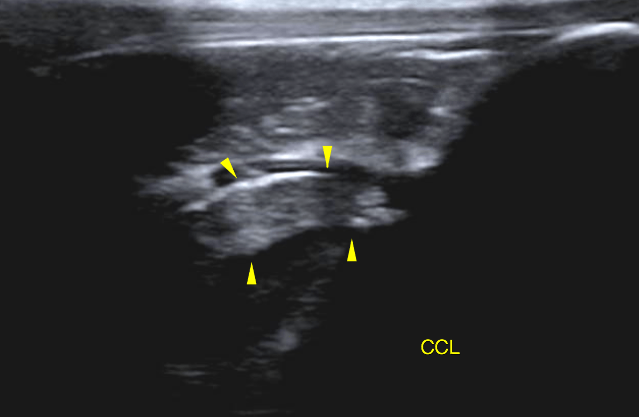
The patient is a 4 year old FS Golden Retriever dog with intermittant lameness of the left hind leg. R/O partial CCL tear left stifle
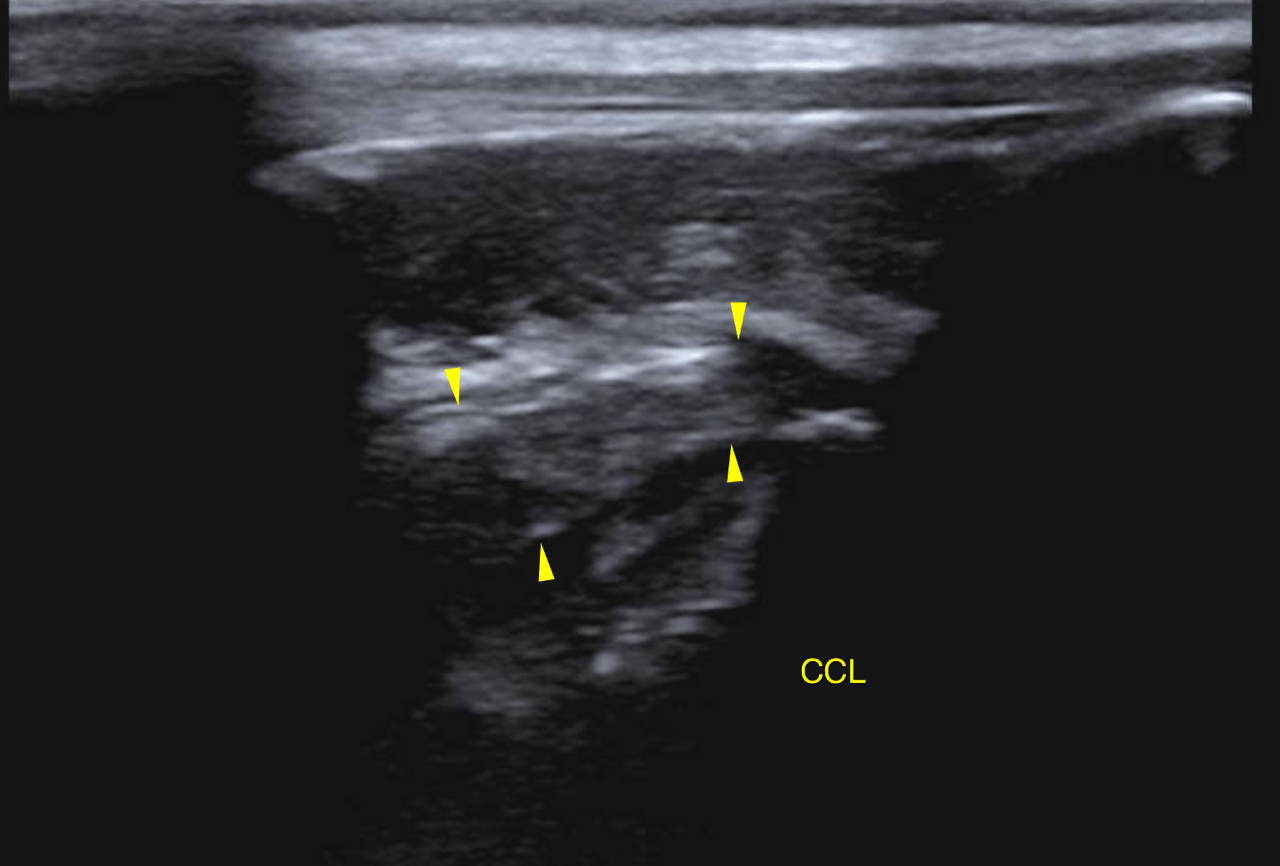
The patient is a 4 year old FS Golden Retriever dog with intermittant lameness of the left hind leg. R/O partial CCL tear left stifle