An 11-year-old SF dog was presented for evaluation of a previously imaged cranial abdominal mass. Physical examination, CBC, and serum biochemistry were all within normal limits.
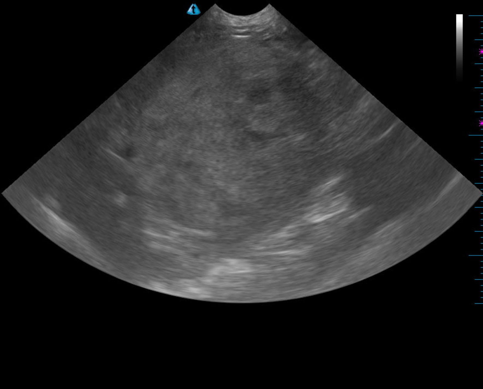
Left sided liver mass. The mass is approximately the same size at +/- 8.0 cm without surrounding inflammation. The lack of growth would suggest hepatoma or a relatively benign process. However, the more malignant factor lies in the potential for lobar torsion, which is life threatening. Therefore, resection is recommended. Chest radiographs are warranted prior to surgery.
The liver in this patient presented a microcavitated mass that occupied the left liver. The mass measured approximately +/- 8.0 cm, which is approximately the same size without surrounding inflammation. This was connected to the cranial aspect of the left liver by a bridge of approximately 3.0 cm. This appeared to be subjectively isolated. The gallbladder and right liver appeared structurally unremarkable.
None
Information about and images from the previous ultrasound are not provided.
Cyst/neoplasia/granuloma/abscess – liver, spleen, lymph node, mesentery, kidney
Hematoma
Hydronephrosis
None