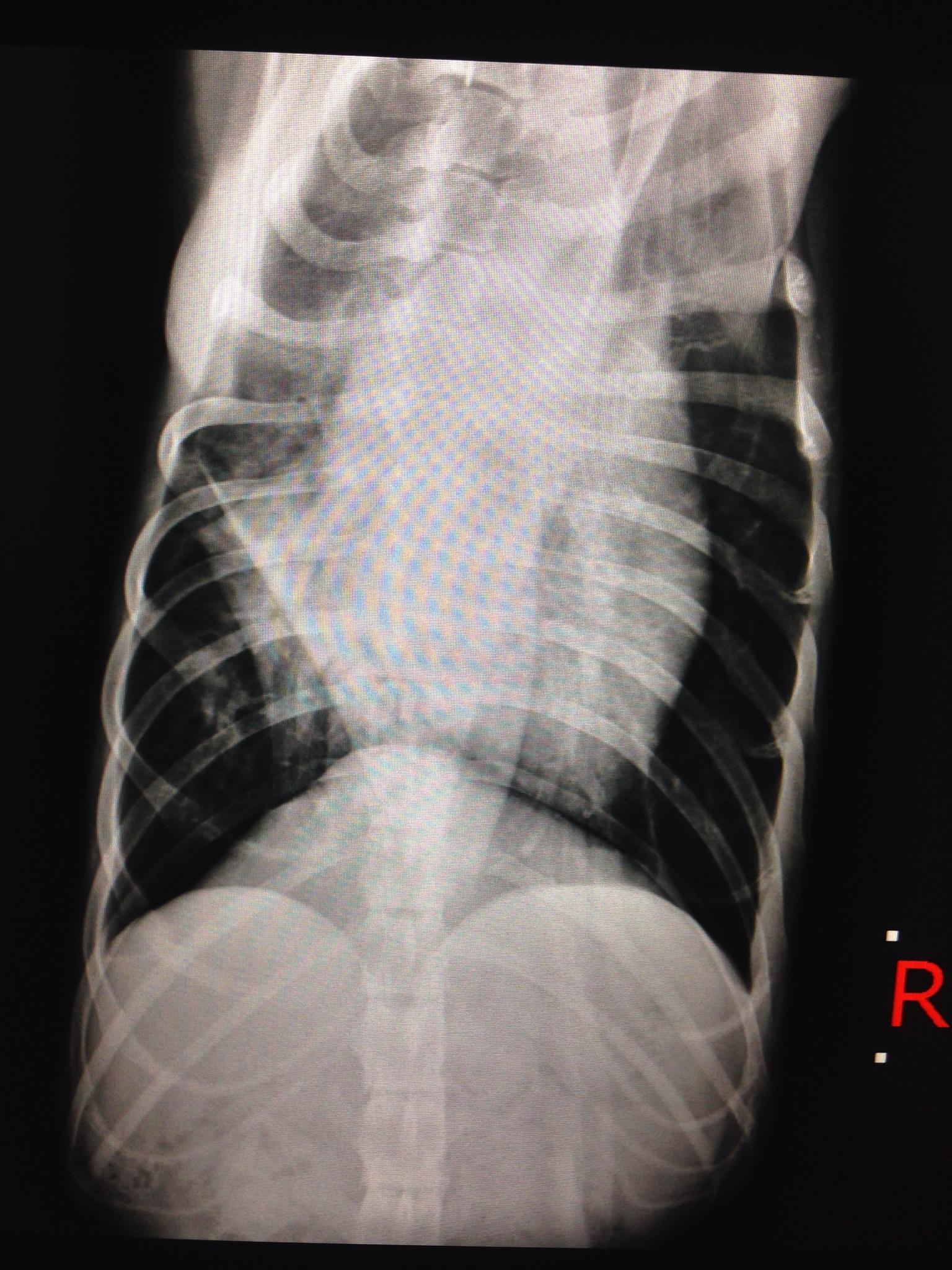
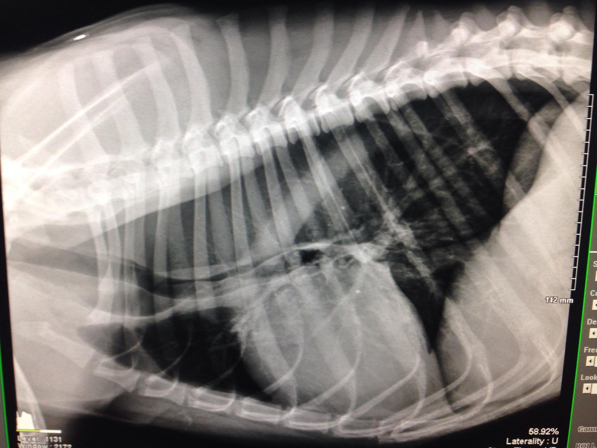
A 3-year-old SF German shepherd was presented for evaluation of a grade IV/VI murmur and normal blood pressure. Survey thoracic radiographs showed thoracic megaesophagus with aspiration pneumonia. On ECG, sinus arrhythmia was evident.
A 3-year-old SF German shepherd was presented for evaluation of a grade IV/VI murmur and normal blood pressure. Survey thoracic radiographs showed thoracic megaesophagus with aspiration pneumonia. On ECG, sinus arrhythmia was evident.
Subaortic Stenosis – severe, concentric hypertrophy present
Aortic Regurgitation – mild
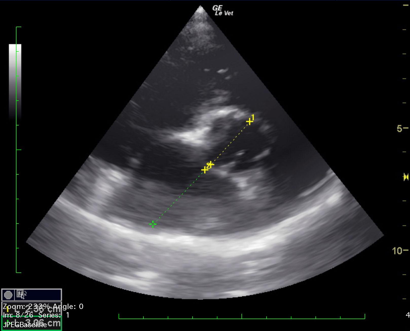
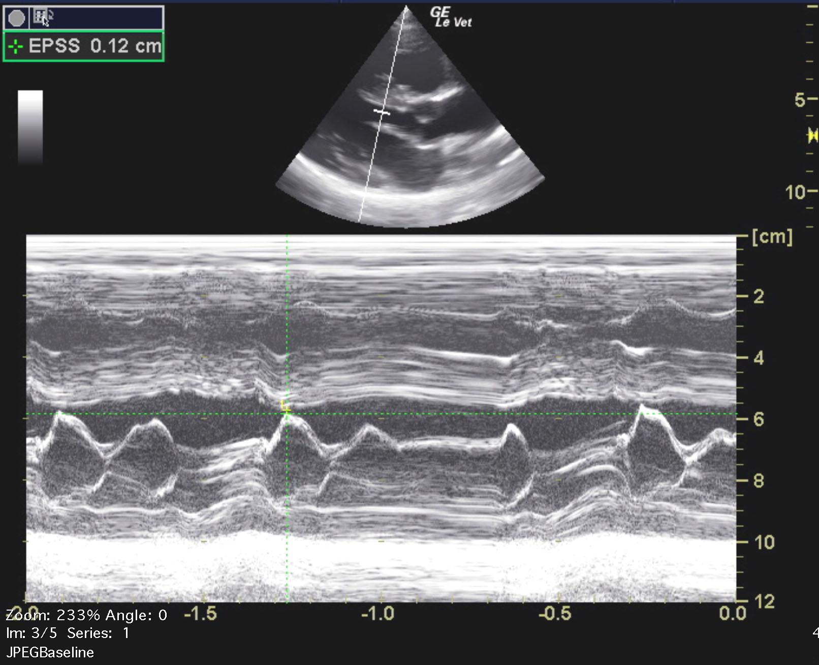
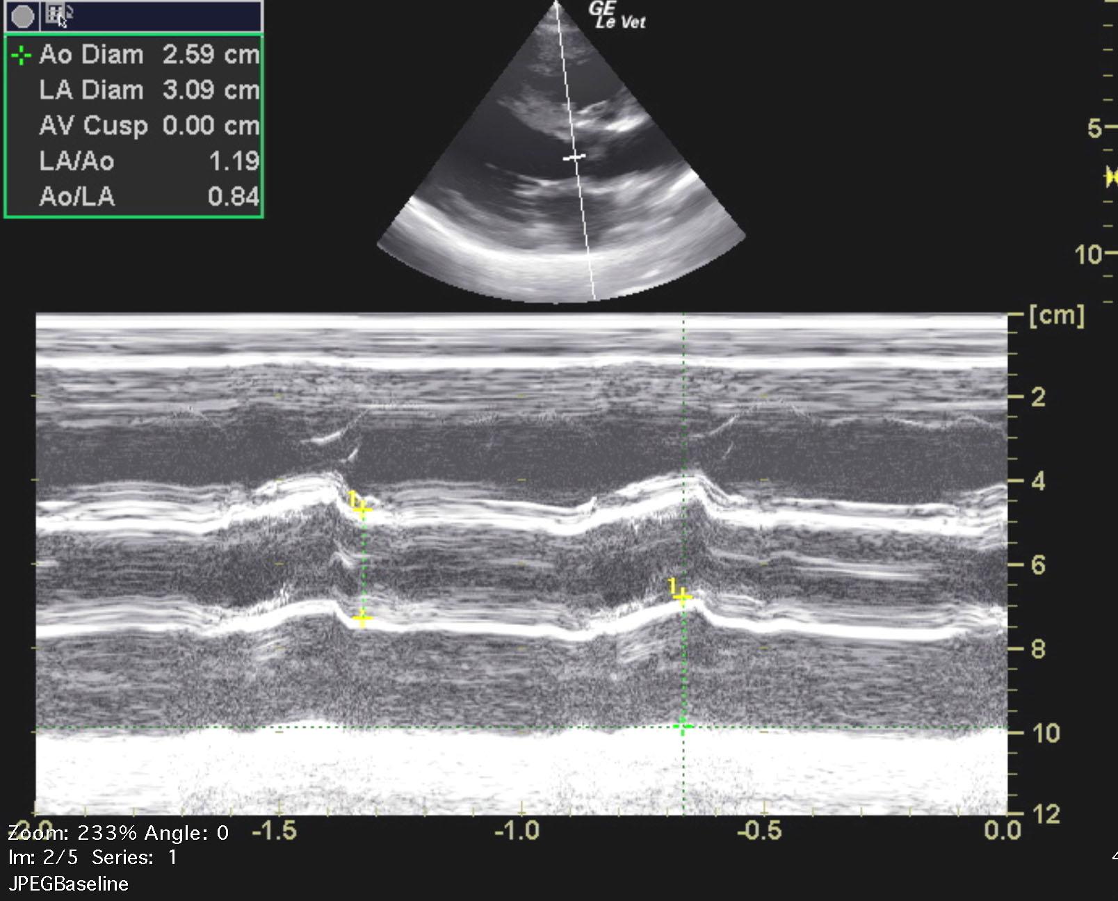
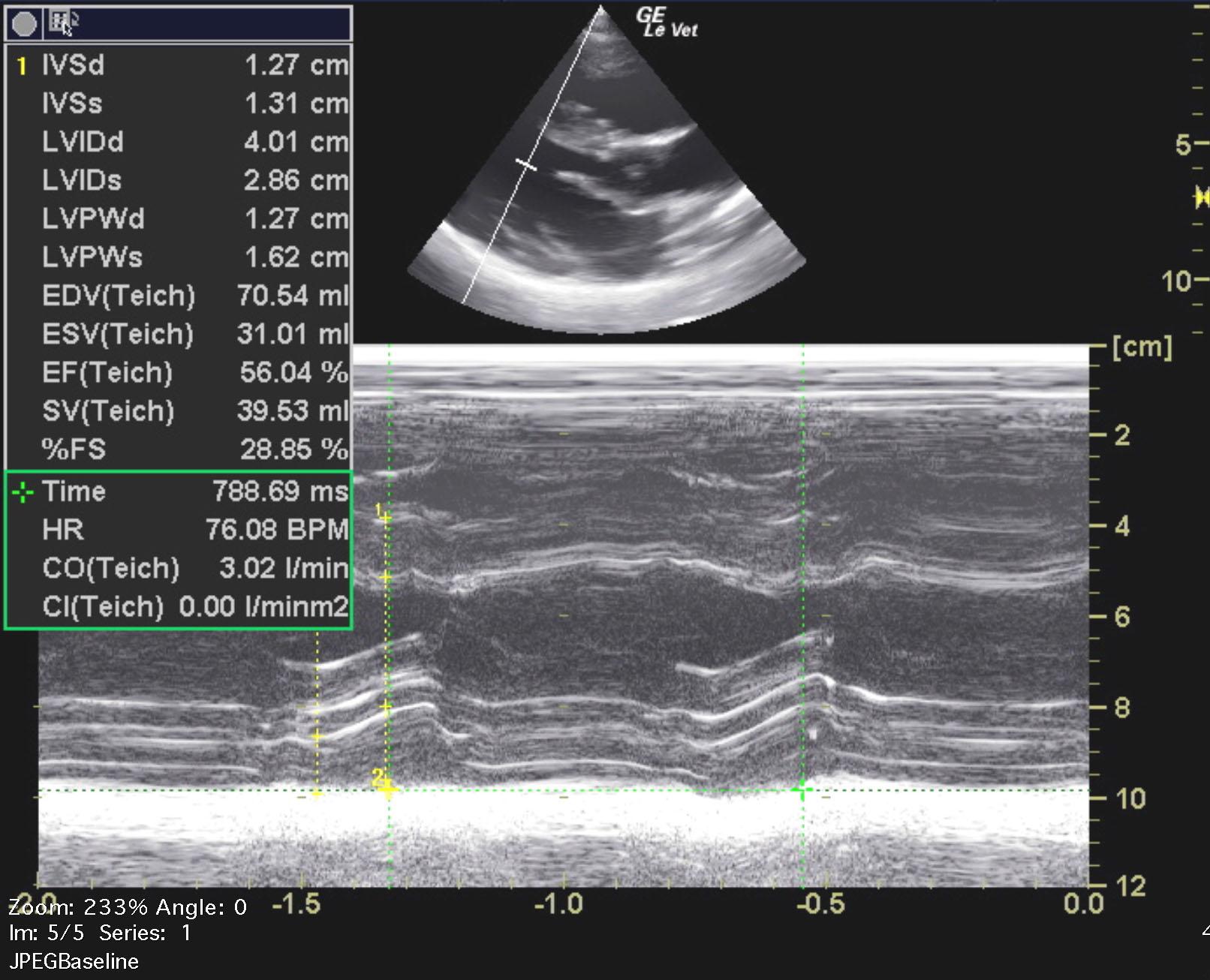
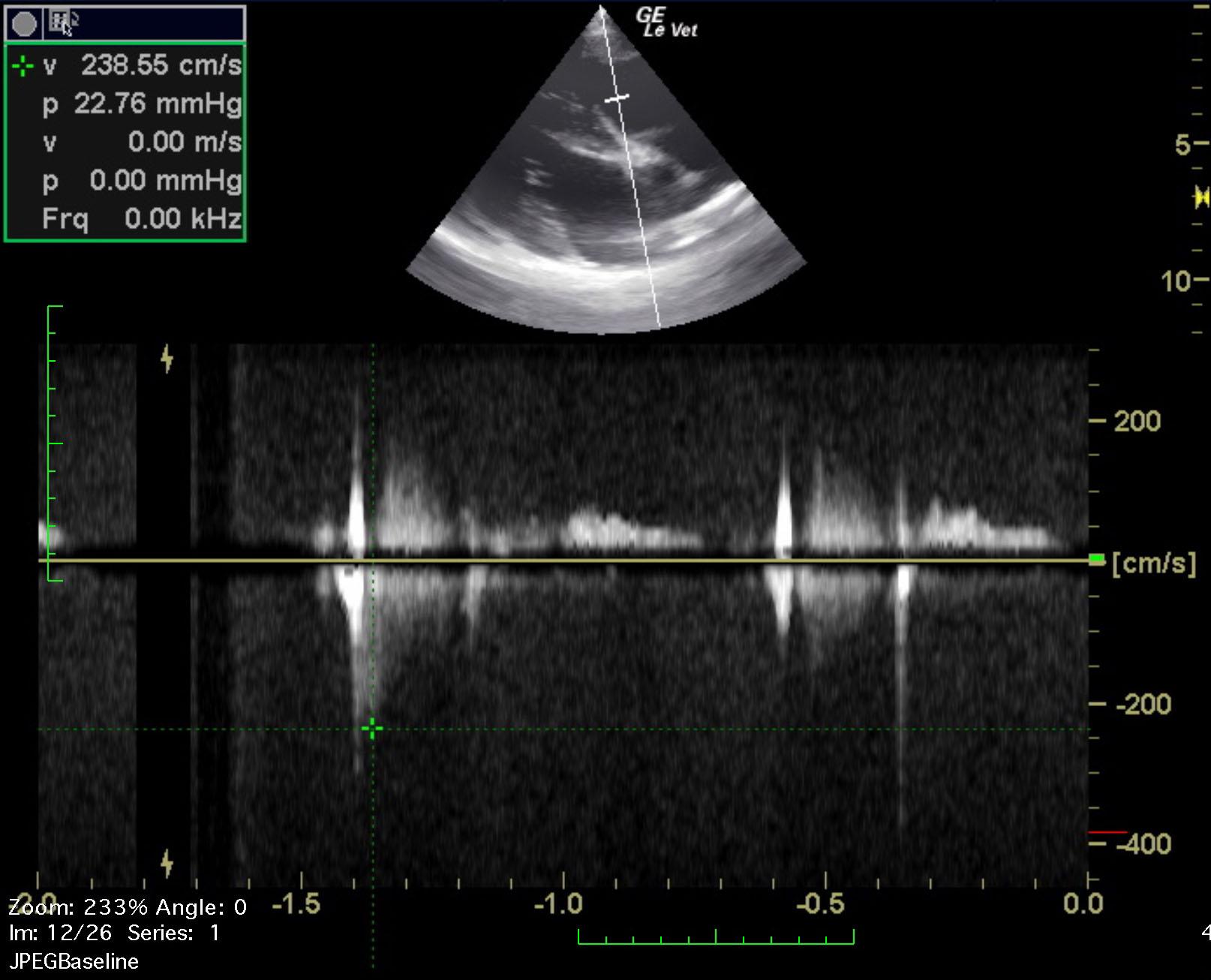
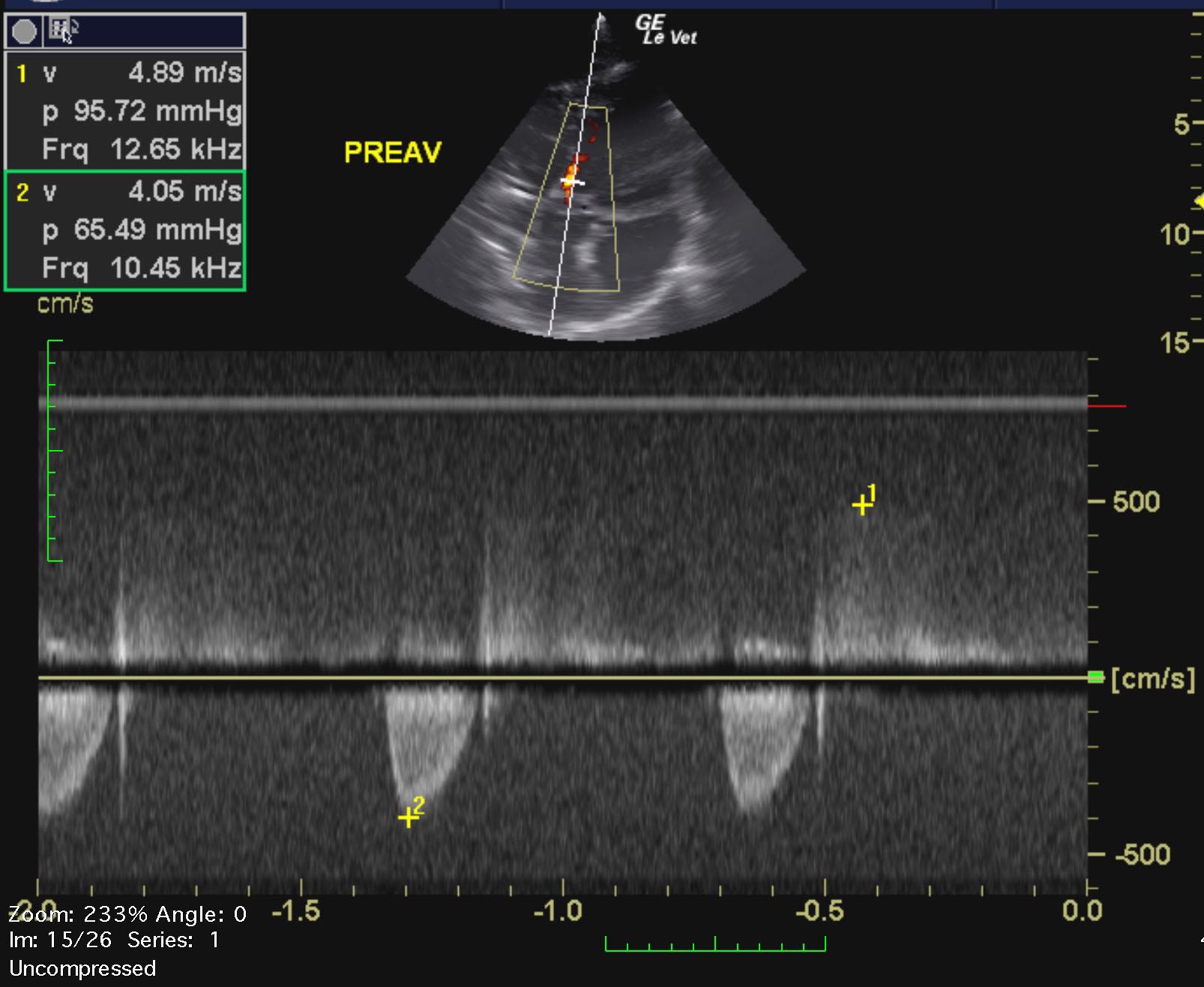
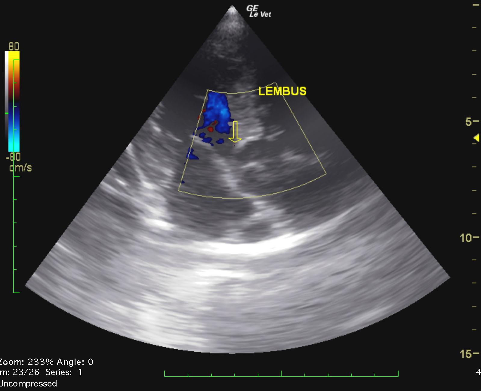
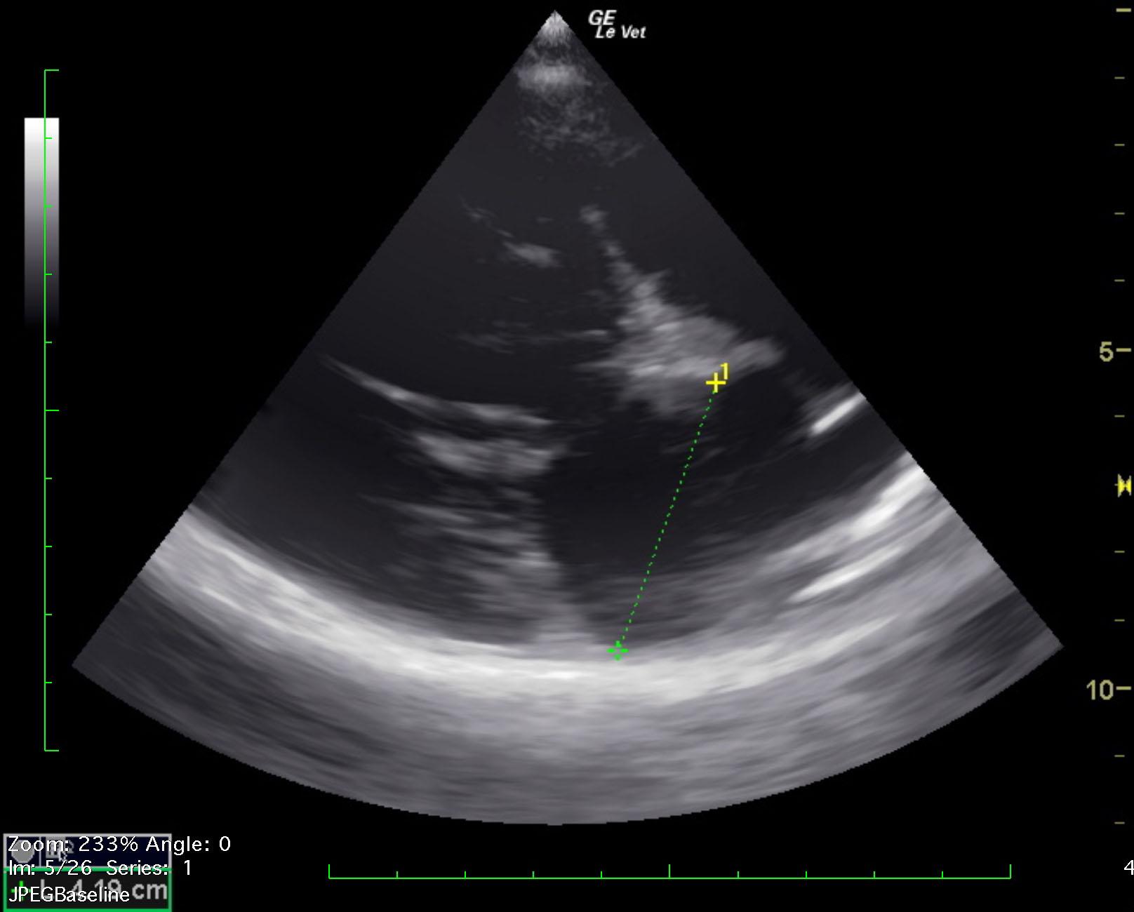
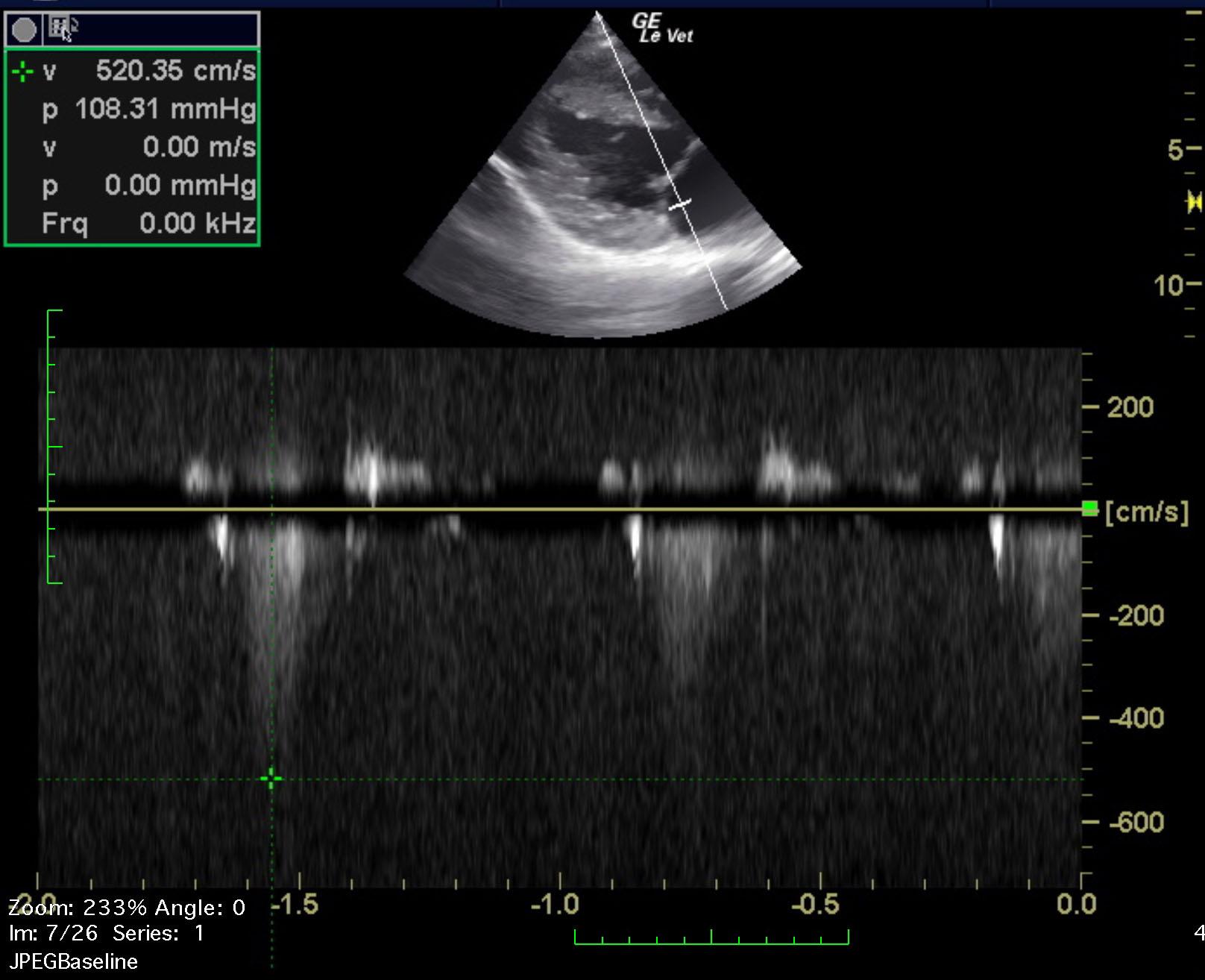
The left ventricular chamber measures normal in size. The wall thicknesses are mildly hypertrophied. There is normal systolic function. The left atrium is normal in size (La:Ao 1.30). The visible right heart is normal in size. There is a notable subaortic ridge. Color Doppler identifies an increase in flow and turbulence associated within the suboartic ridge/LVOT. The aortic valve is mildly thickened in some views/grossly normal in other views. The peak velocity of aortic outflow is consistent with severe SAS (LVOT 4.89m/s = 96mmHg). Mild aortic regurgitation is visualized. The mitral valve may be minimally thickened and there is mild MR (peak velocity ~5.20m/s). All other valves appear normal. There is normal, laminar flow across the RVOT/PA (RVOT/PA 1.24m/s). No effusions or masses visualized. IVDd – 1.27cm LVIDd – 4.01cm LVFWd – 1.27cm IVDs – 1.31cm LVIDs – 2.86cm LVFWs – 1.62cm FS – 29%.
None
Cardiac – congenital heart disease (aortic stenosis, PDA, pulmonic stenosis, atrial/septal defect), vegetative endocarditis, dilated cardiomyopathy
Congenital/idiopathic megaesophagus
None