A 7-year-old NM beagle was presented for evaluation of hematuria and stranguria. Urinalysis showed SG of 1.014 and mild proteinuria.
A 7-year-old NM beagle was presented for evaluation of hematuria and stranguria. Urinalysis showed SG of 1.014 and mild proteinuria.
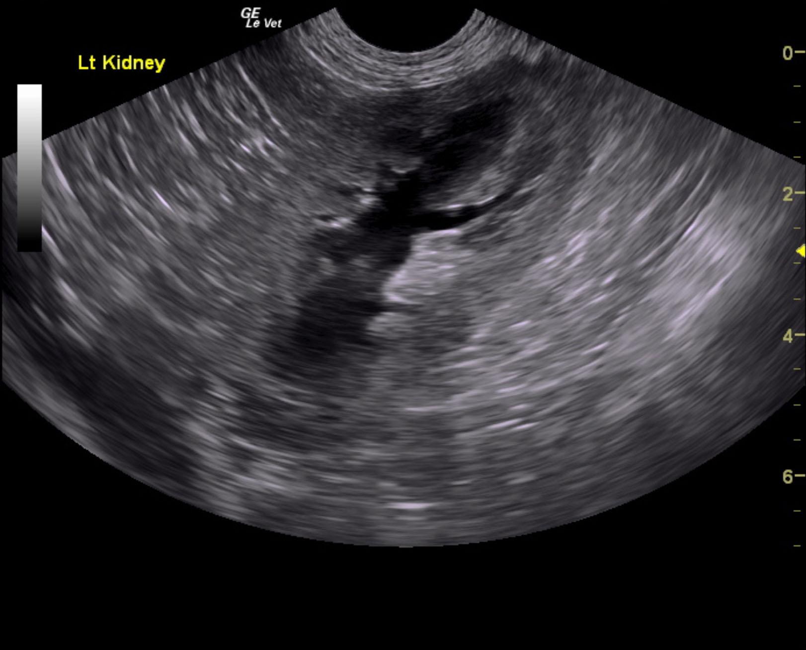
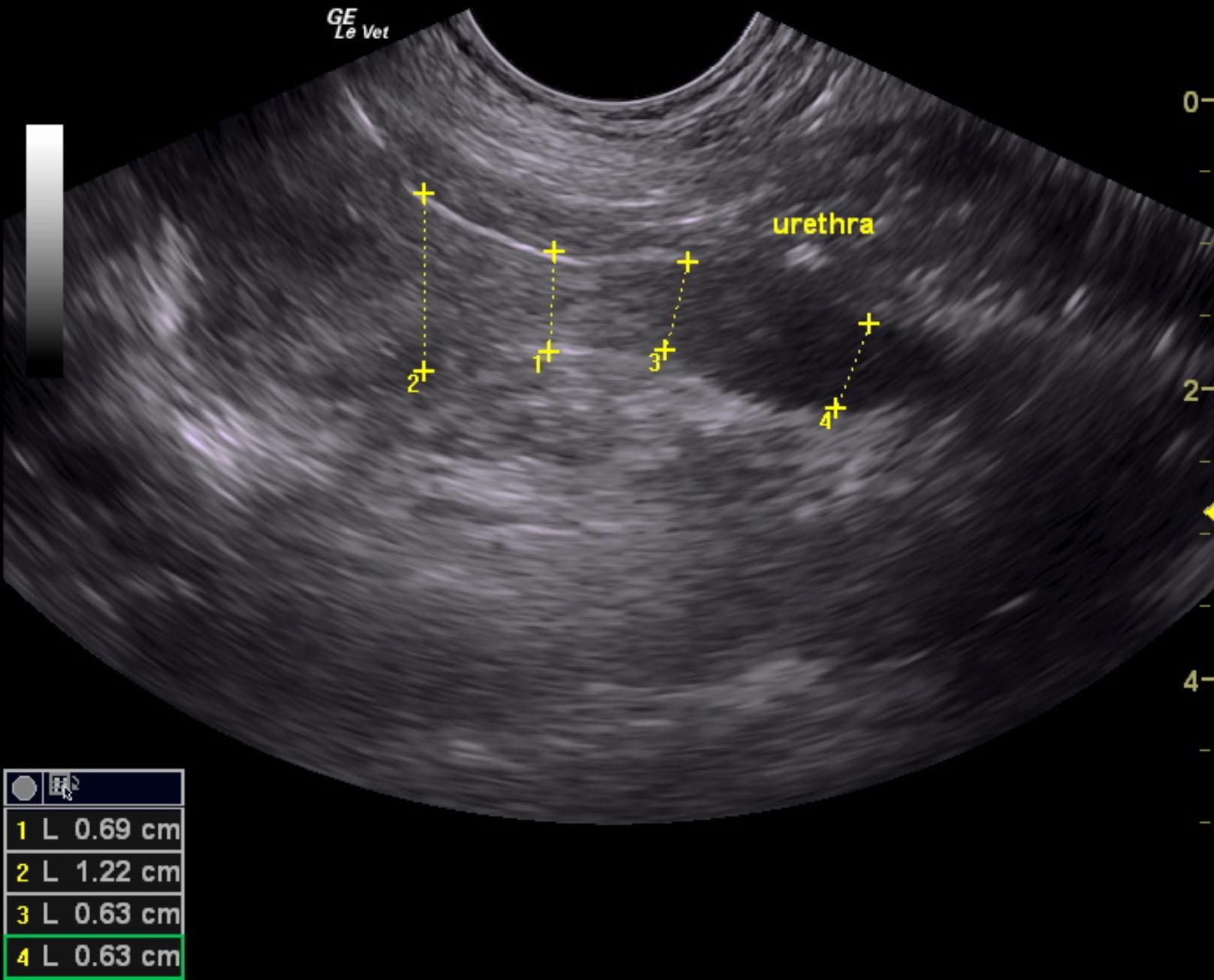
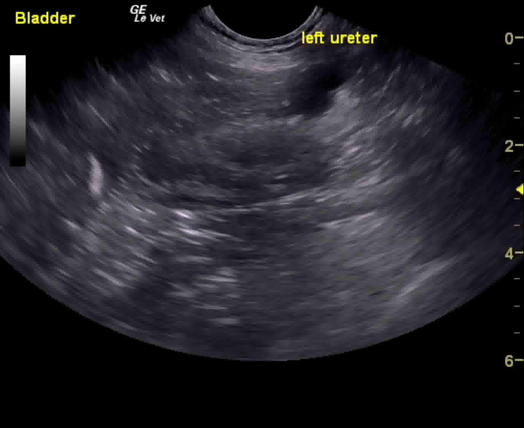
Urethral and trigonal mass with left ureteral obstruction and mild to moderate hydronephrosis left kidney with pericapsular fluid accumulation.
Referral for stent placement would be recommended in this patient; however, prognosis is guarded. Urethral and left ureteral stents would be necessary in this case with potential surgical ablation of the urethral and trigonal tumor.
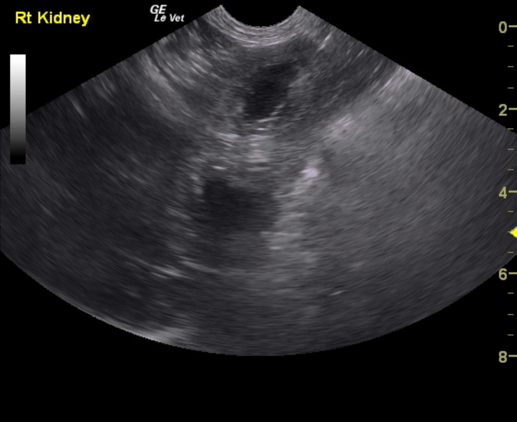
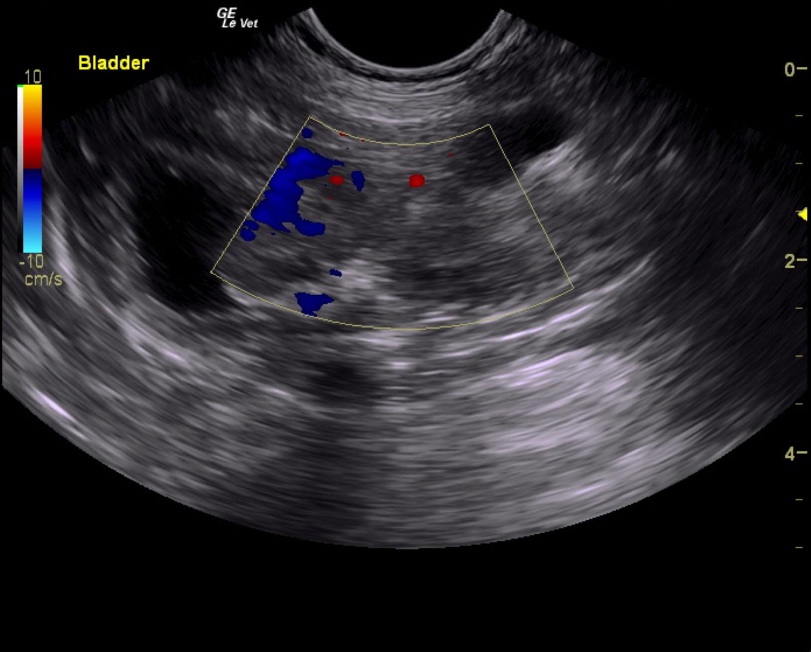
Mild hydronephrosis left kidney noted 1.5 x 0.45 cm. Pericapsular fluid accumulation was noted around the left kidney. Right kidney had mild degenerative changes, normal size and contour. Urinary bladder presented 3 cm+ irregular trigonal and urethral mass. Focal areas of mineralization noted. Apical polypoid changes were noted in the bladder as well. Color flow Doppler revealed moderate positive signals to the mass itself. The pelvic urethra was invaded at least 4 cm from the cystourethral junction to a width of 1.22 cm to 0.63 cm. Left ureter was obstructed owing to the mass, approximately 1 cm hydroureter noted.
None
Bladder – neoplasia, bacterial cystitis, uroliths, granulomatous cystitis, polyploid cystitis
Prostate – neoplasia, prostatitis, abscess
Urethra – urethritis, neoplasia, lith
None