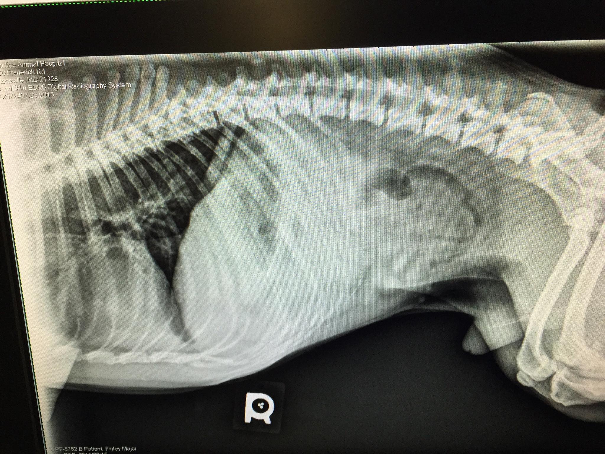
A 4-year-old NM French Bulldog was presented for evaluation of intermittent sub-acute vomiting.A small intestinal obstructive pattern was noted at the ileocecal region and on survey radiographs.
Sonographic Differential Diagnosis
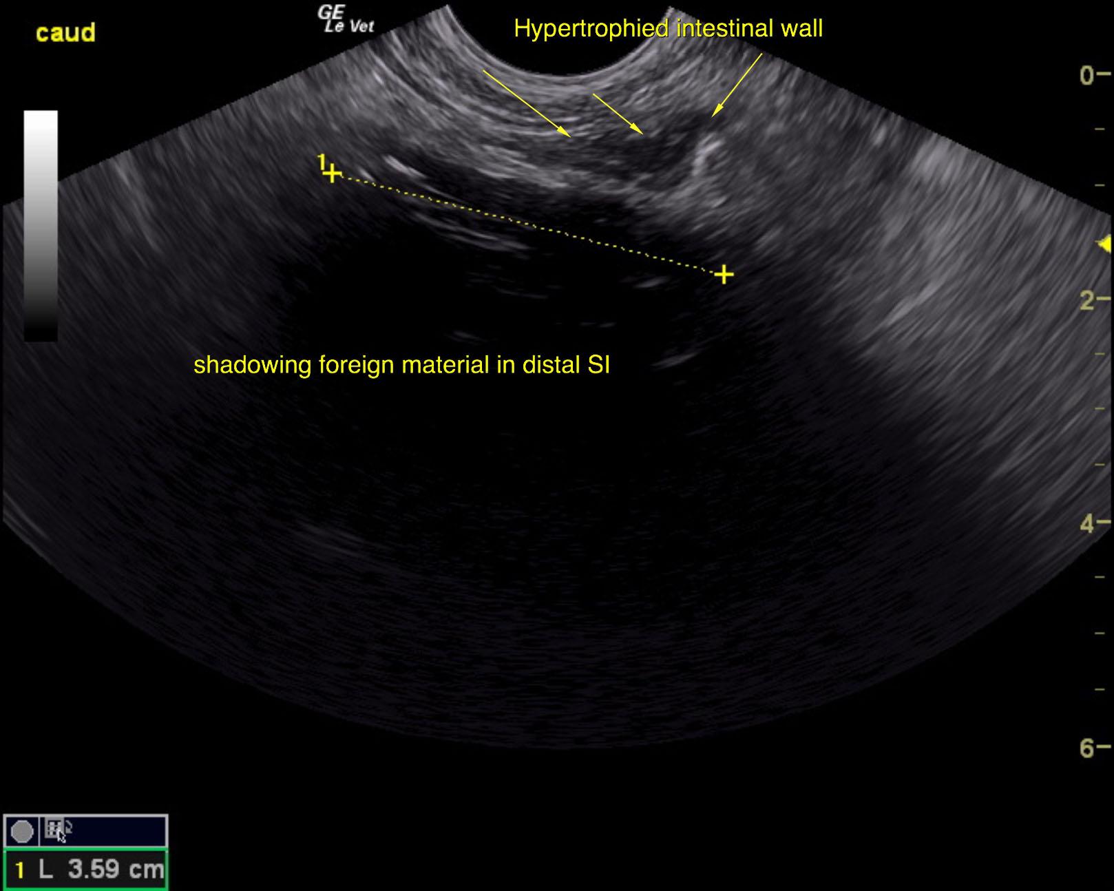
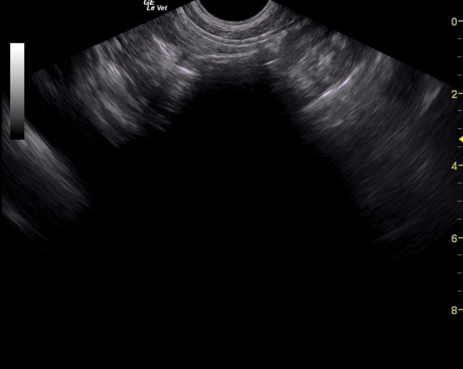
Foreign body obstruction.
Distal small intestine/ileocecal region with hard shadowing material such as wood or similar.
The distal small intestine revealed slight thickening. Biopsies would be ideal in this case at the time of surgery.
Image Interpretation
The caudal small intestine revealed distinctly shadowing material. This is likely fluid absorbing. The descending colon was unremarkable. Mesenteric lymph node measured 2.36 cm and normal in length x width ratio. The mesenteric lymph node was mildly hypoechoic.