A 7-year-old NM Scottish terrier was presented for evaluation of hematuria that had initially responded to cortisone therapy. Urinalysis showed SG of 1.008, mild proteinuria, and hematuria. Survey radiographs were within normal limits.
Sonographic Differential Diagnosis
Bladder mass
Image Interpretation
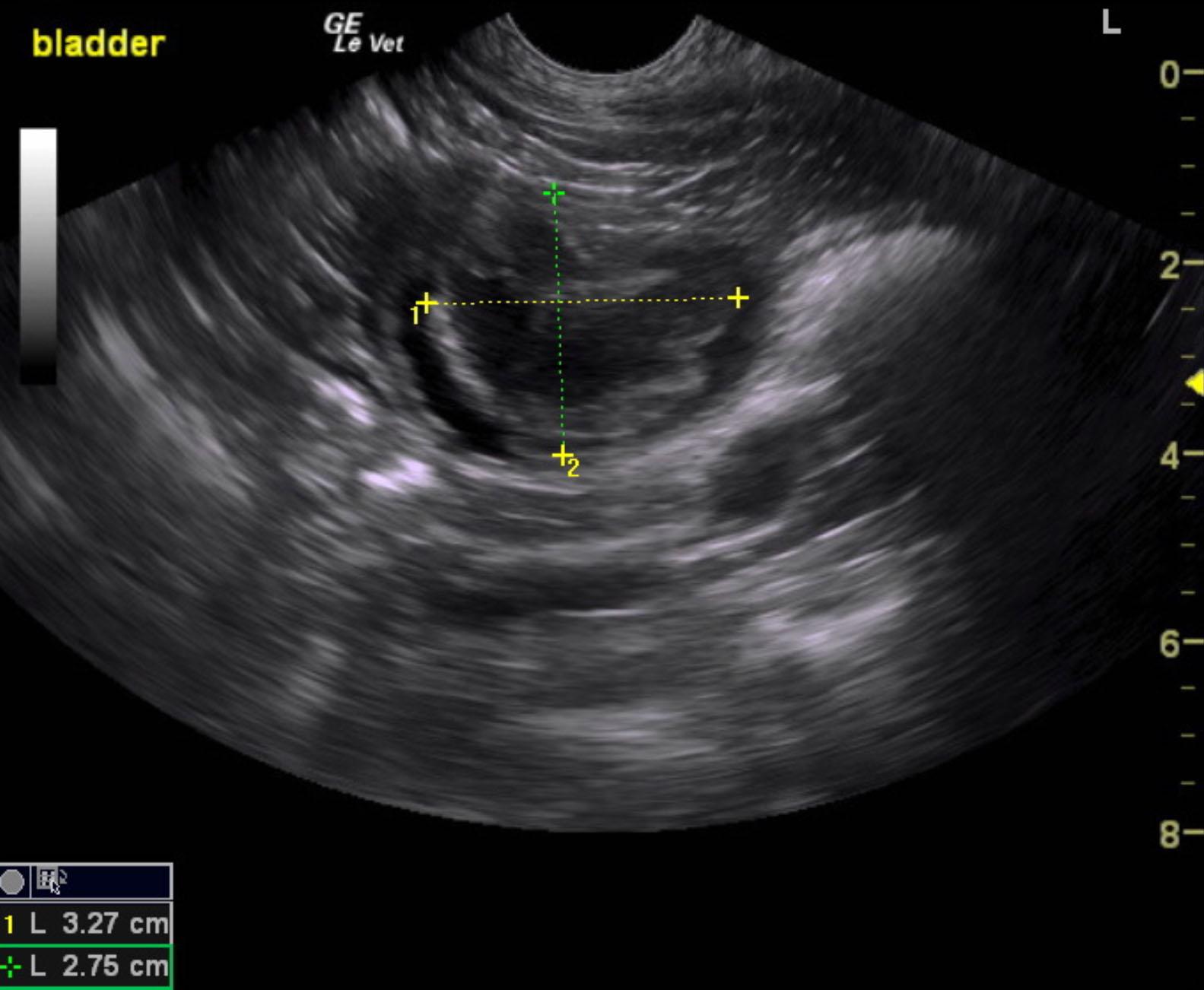
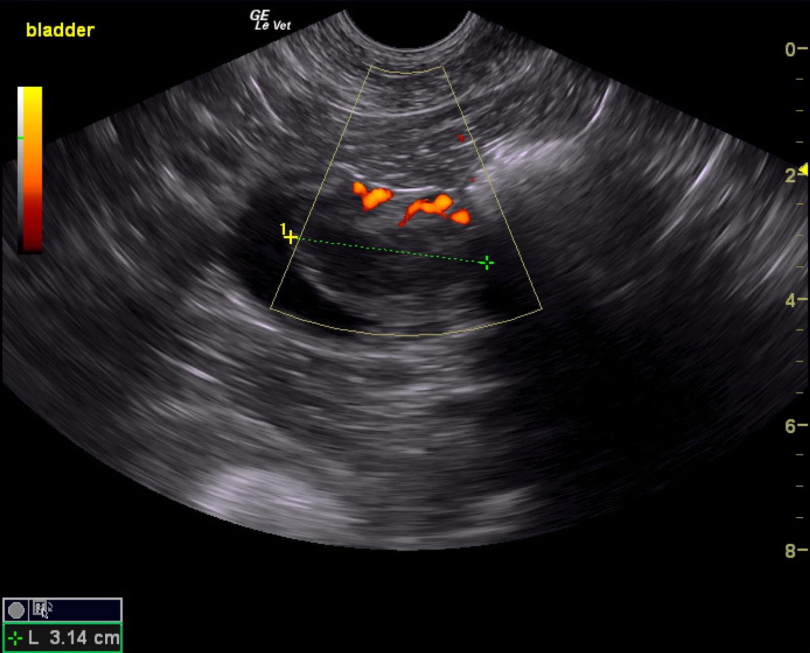
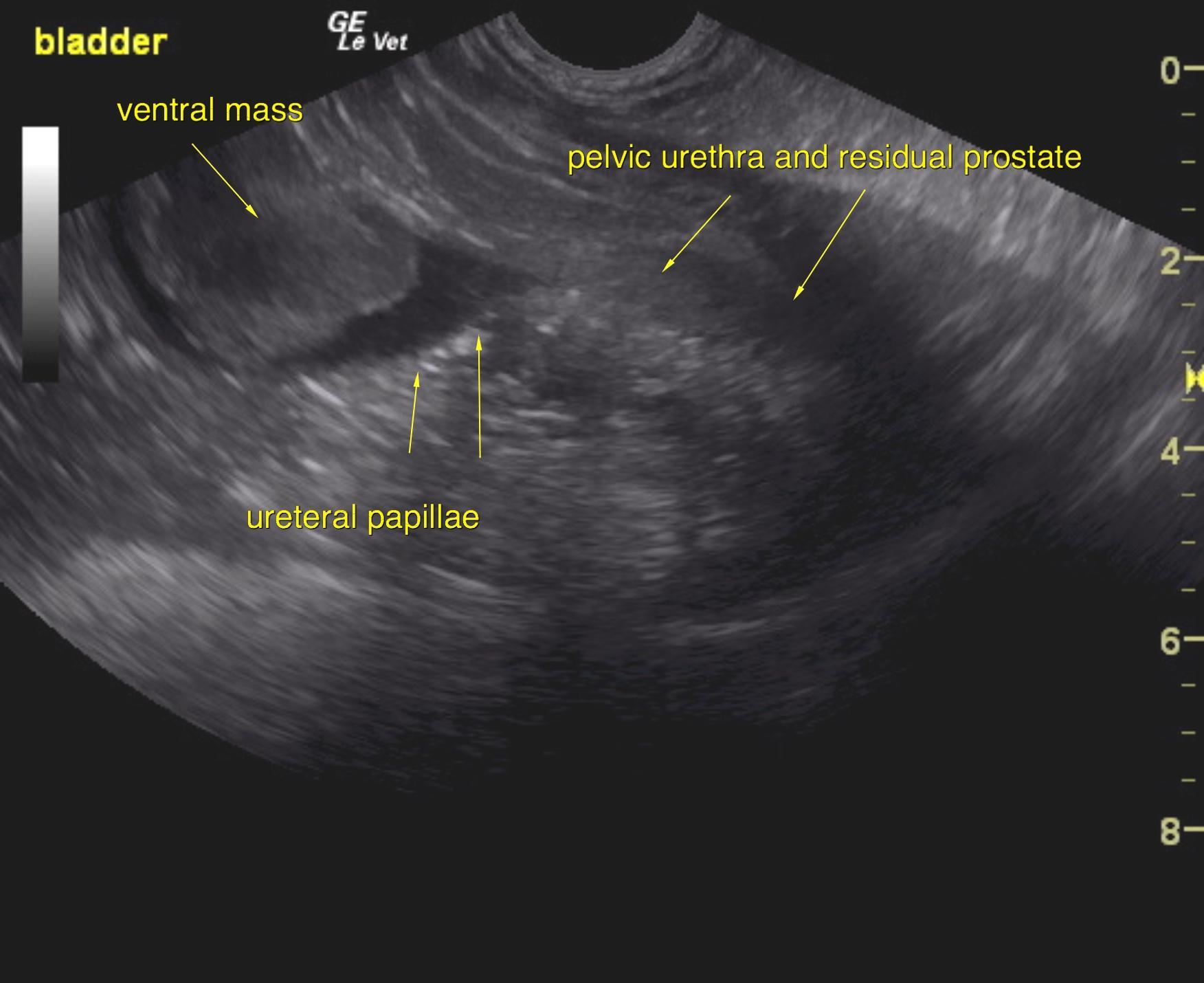
The urinary bladder presented a 3.0 cm mass that was deriving from the ventral wall approximately 1.0 cm cranial to the cystourethral junction. The mass at its maximum width measured 3.27 x 2.75 cm. The mass was significantly vascular on power Doppler. The trigone, pelvic urethra and prostate all appeared free of evident pathology.