08_00153 Moose E Splenic neoplasia

Case Study

08_00153 Moose E Splenic neoplasia

A 7-year-old NM Mastiff mix was presented for evaluation of decreased appetite, weight loss, vomiting, and lethargy. Splenomegaly was present on physical examination. Abnormalities on CBC and serum biochemistry were mild anemia, thrombocytopenia, and elevated liver enzyme activity. Pulmonary nodules were evident on survey thoracic radiographs.
 

Sonographic Differential Diagnosis

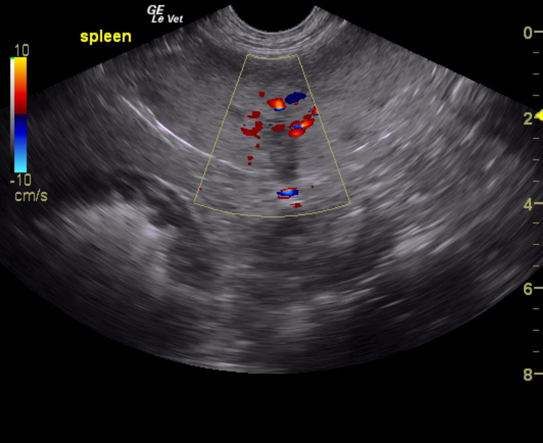
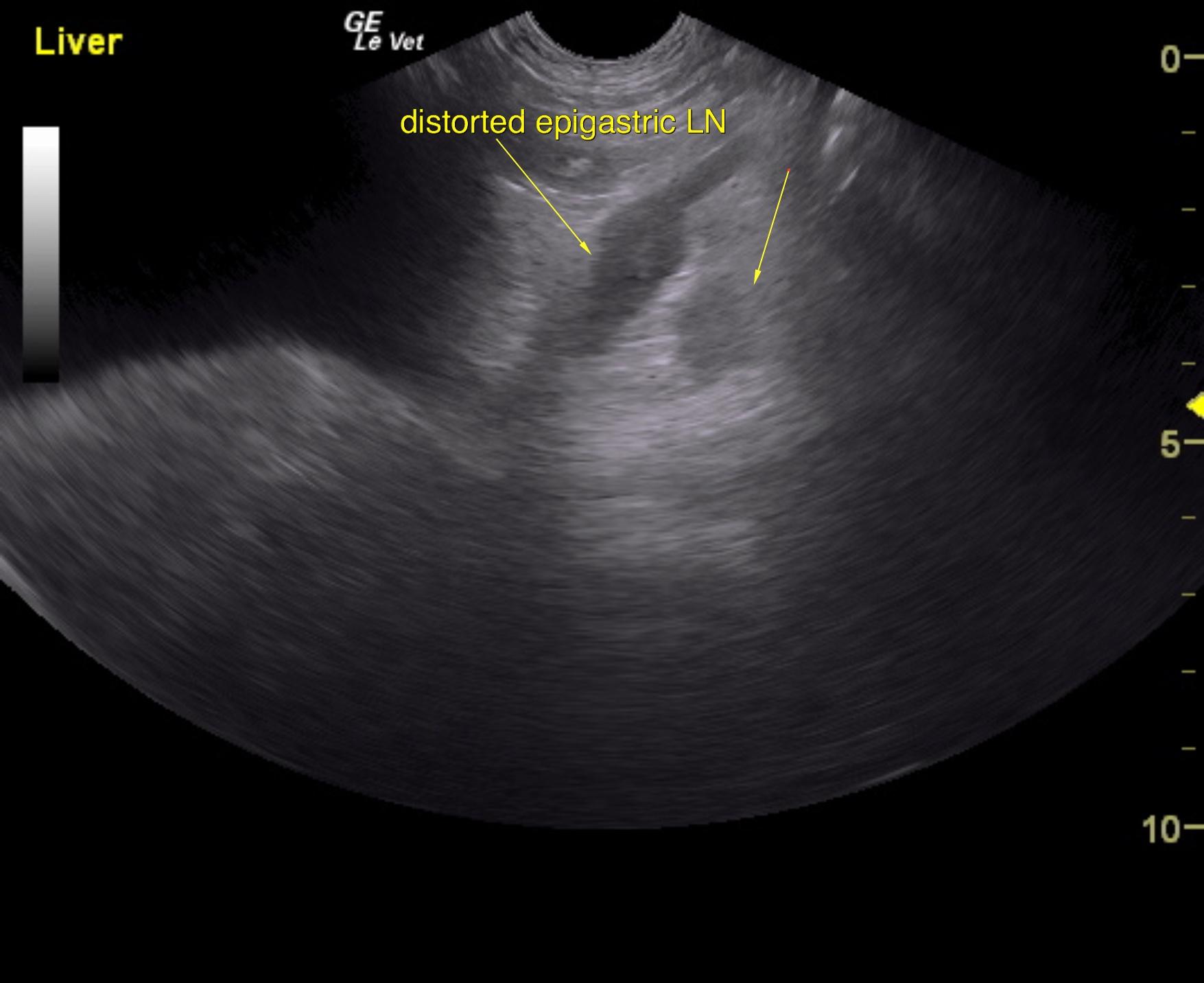
Splenic neoplastic pattern, likely involving epigastric lymph node and liver. Ultrasound-guided FNA of the spleen, epigastric lymph node and liver would be recommended.
Potential early metastatic disease to the chest.
Concurrent splenic thrombosis owing to hypercoagulable state.

Image Interpretation

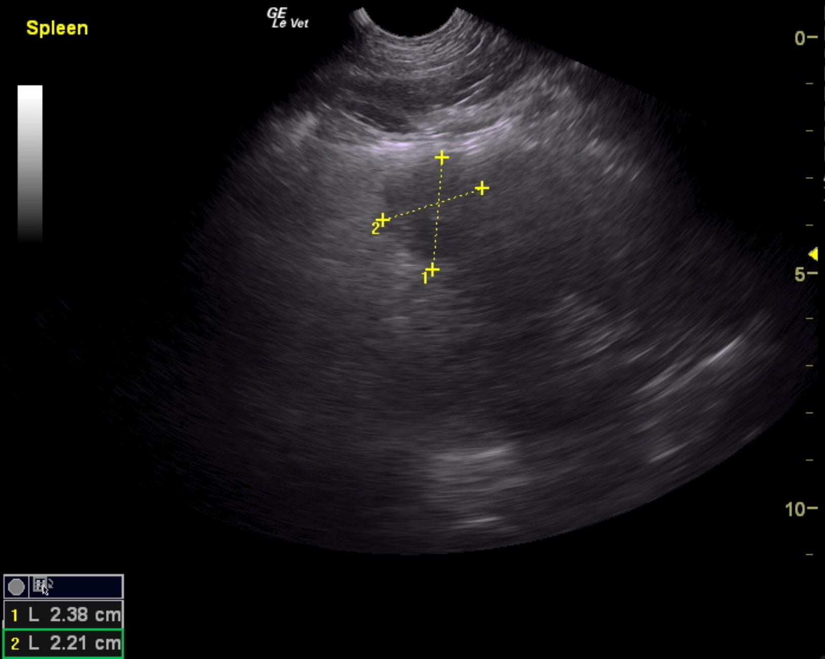
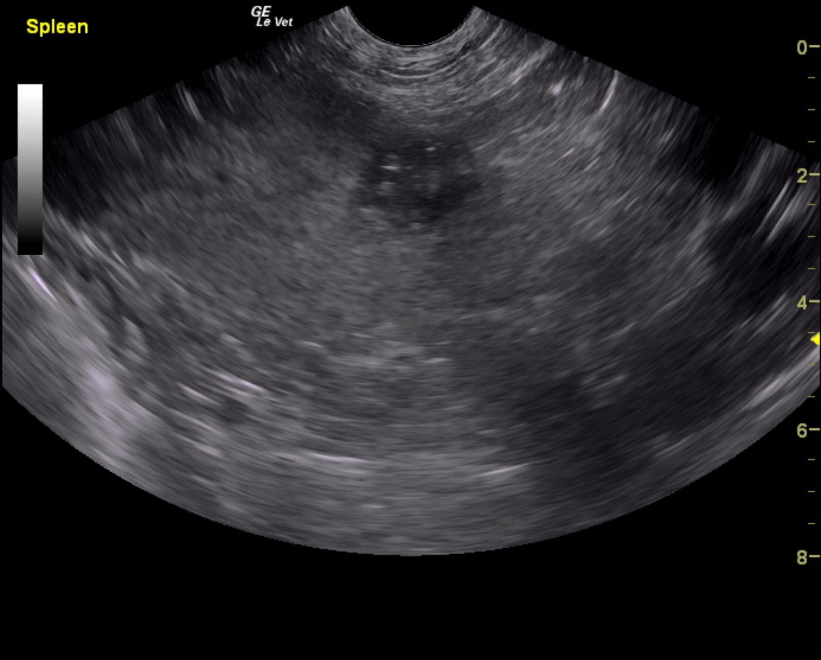
The spleen revealed multiple, hypoechoic target type nodules. One of which measured 2.38 x 2.21 cm. Multiple splenic nodules revealed loss of architecture and multi focal hyperechoic changes. Splenic thrombus was noted.
A mixed, hypoechoic distorted lymph node was noted in the epigastric lymph node region measuring approximately 2.5 x 2.0 cm with pericapsular inflammatory pattern.

DX

Suspect splenic neoplasia

Outcome

None

Clinical Differential Diagnosis

Metastatic splenic neoplasia – hemangiosarcoma, sarcoma
Fungal disease

Sampling

None

Patient Information

Gender : Male, Neutered
Species : Canine
Type of Imaging : Ultrasound
Status : Complete

Clinical Signs

  • Anorexia
  • Lethargy
  • Vomiting
  • Weight loss

Exam Finding

  • Splenomegaly

Images

mosse1mos2mos3mos4

Blood Chemistry

  • Elevated Liver Enzymes

CBC

  • Platelet Count, Low
  • RBC, Low

Clinical Signs

  • Anorexia
  • Lethargy
  • Vomiting
  • Weight loss