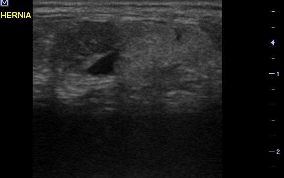
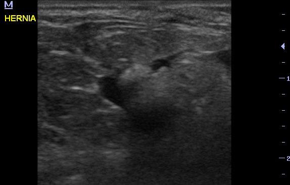
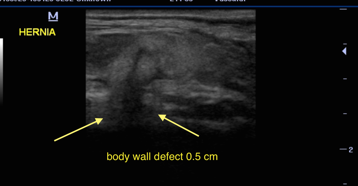
A 3-year-old SF Dachshund that had developed a small lump midline after being spayed at 6 months of age was presented for evaluation of the lump growing larger and firmer.
Sonographic Differential Diagnosis
Umbilical or body wall hernia at midline. This is surgically rectifiable with debridement and closure of the body wall at the linea alba.
Image Interpretation
A small umbilical hernia was noted in this patient with extrusion of approximately 1.5 x 1.0 cm of fat. Minor body wall irregularity was noted. The hernia measured approximately 0.5 cm in width. Minor inflammatory pattern was noted associated with the hernia and minor fluid accumulation.