03_00451 Zoey L PSS

Case Study

03_00451 Zoey L PSS

 A 2-year-old SF Pug was presented for evaluation of intermittent vomiting, anorexia, and lethargy; with biochemistry showing elevated bile acids. Physical examination and CBC were both normal. The only finding on urinalysis was isosthenuria. Abnormalities on serum biochemistry were elevated AST (89) and ALT (276) activity, and low BUN (1.8).
Sonographic Differential Diagnosis

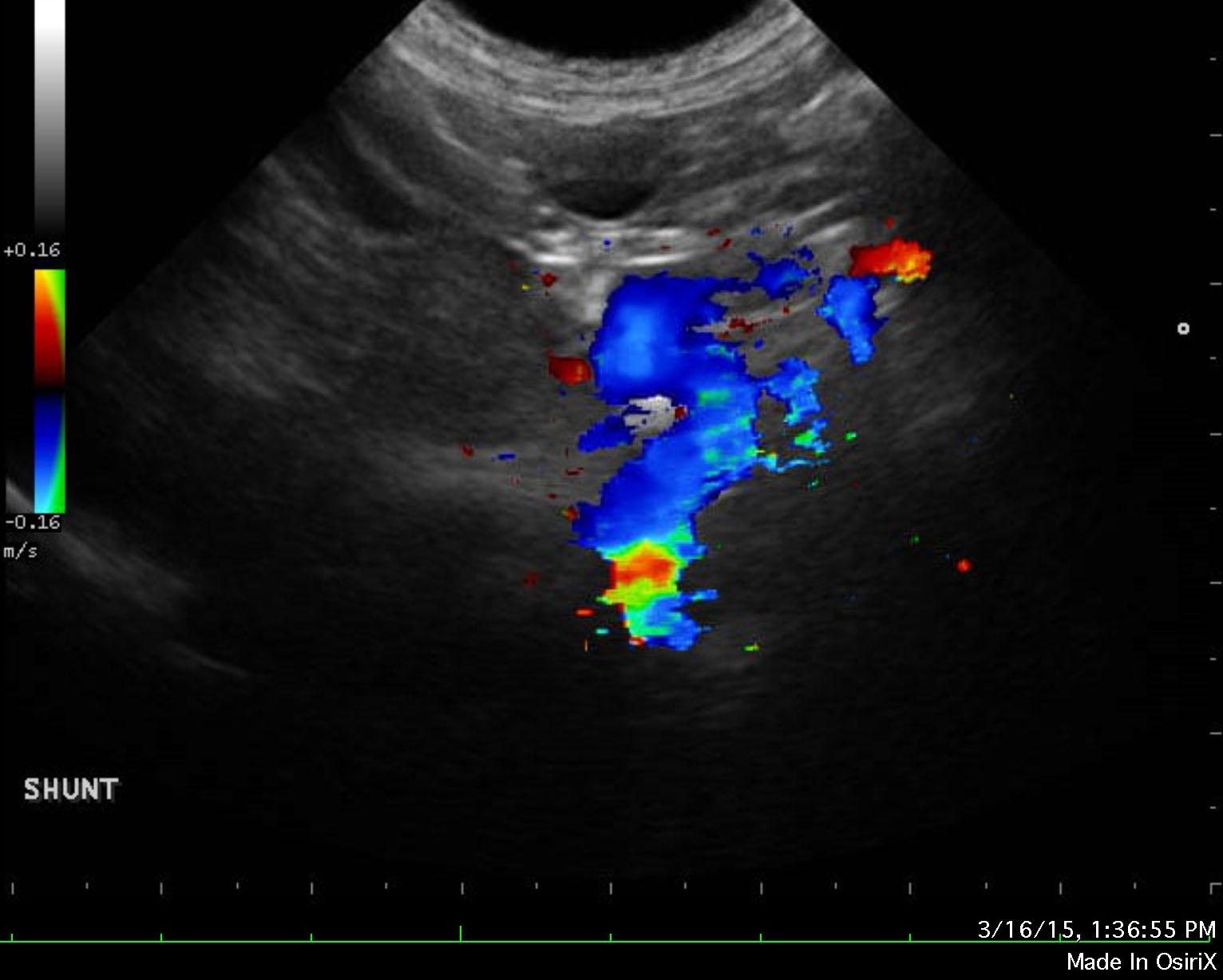
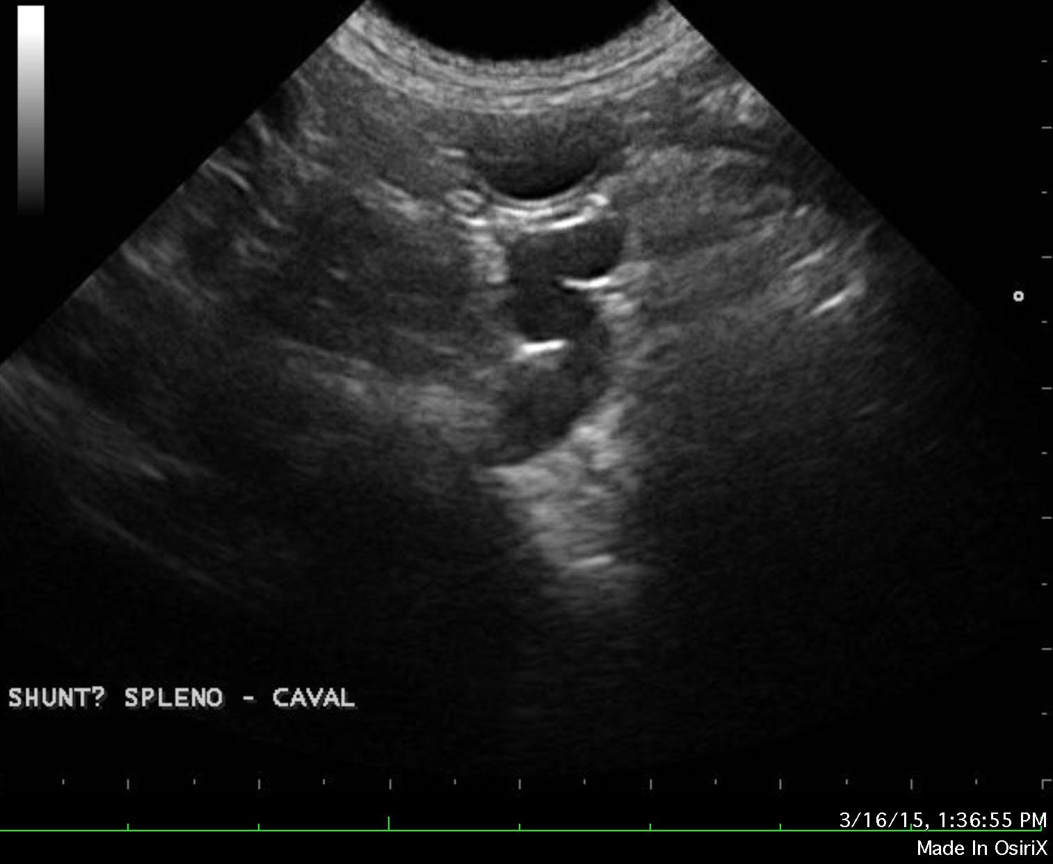
Suspect splenoazygos shunt. Possible splenocaval shunt, yet ratio is most consistent with splenoazygos shunt. The turbulent flow in the vena cava may be color bleeding from adjacent turbulent flow within the suspected splenoazygos shunt.
Severe microhepatica.
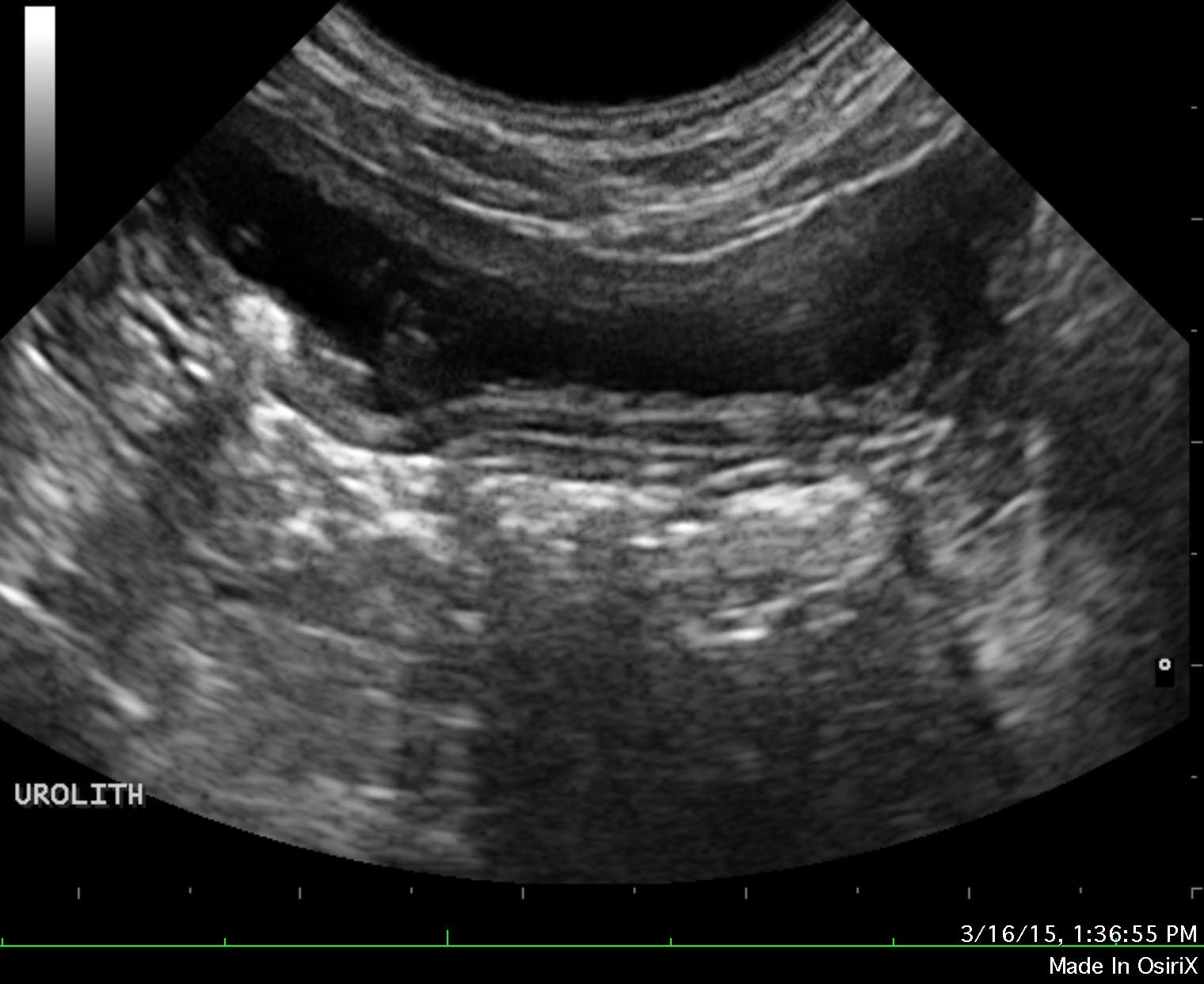
Bladder sand.

Image Interpretation

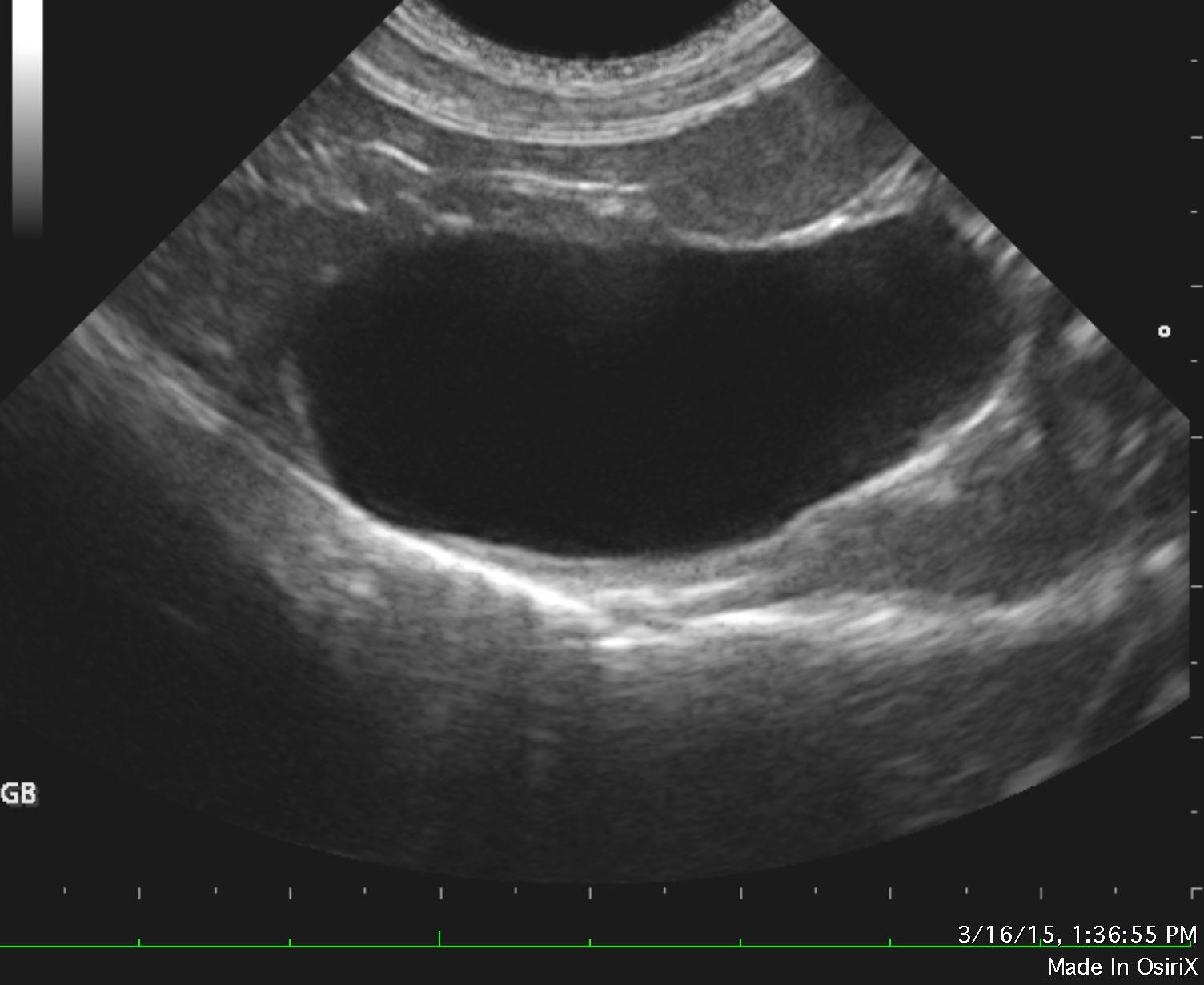
The liver revealed significant microhepatica. The portal vein at the level of the aortic hiatus measured 0.53 cm, vena cava 0.5 cm. At the level of the portal hilus the portal vein measured 0.36 cm and vena cava 0.91 cm. There is a tortuous vessel coursing dorsally and appears to enter into the aortic hiatus. Given that the vena cava to aorta ratio appears to be 1:1, this would be consistent with splenoazygos shunt. The shunt measured 0.8 cm in width and 2.0-4.0 cm in length as it connected the splenic vein junction to the portal vein with the vena cava. The liver presented mildly increased portal markings. The gallbladder was unremarkable and appeared overwhelming in comparison to the subnormal liver size. The common bile duct measured 0.22 cm and was normal. The urinary bladder was mildly thickened with a small amount of sand accumulation measuring 0.5 cm as a grouping.

DX

Portosystemic shunt

Outcome

None

Comments

Ameroid constrictor placement would be recommended. Concurrent portal vein hypoplasia may also be an issue in this patient given the severe microhepatica. I am concerned for potential portal hypertension post surgery. This should be monitored, ideally with intraoperative ultrasound measuring portal vein velocities after either cellophane tie-off or ameroid constrictor placement. Postoperative portal vein velocity measurements are recommended one day and 5 days post surgery to ensure portal hypertension does not develop in this case. Cystotomy for sand removal can be considered, but it is a minor amount and may flush out with fluid therapy. 


Clinical Differential Diagnosis

Liver – porto-caval shunt, primary portal vein hypoplasia, congenital fibrosis, chronic-active hepatitis, granulomatous disease

Sampling

None

Patient Information

Gender : Female, Spayed
Species : Canine
Type of Imaging : Ultrasound
Status : Complete

Clinical Signs

  • Anorexia
  • Lethargy
  • Vomiting

Images

lauzon_sierra_zoey_splenoazygios_shunt_gblauzon_sierra_zoey_splenoazygios_shunt_2lauzon_sierra_zoey_splenoazygios_shuntlauzon_sierra_zoey_splenoazygios_shunt_ub_sand

Blood Chemistry

  • ALT (SGPT), High
  • AST (SGOT), High
  • BUN low
  • Post-Prandial Bile Acids, High

Clinical Signs

  • Anorexia
  • Lethargy
  • Vomiting

Urinalysi

  • Isosthenuria Present