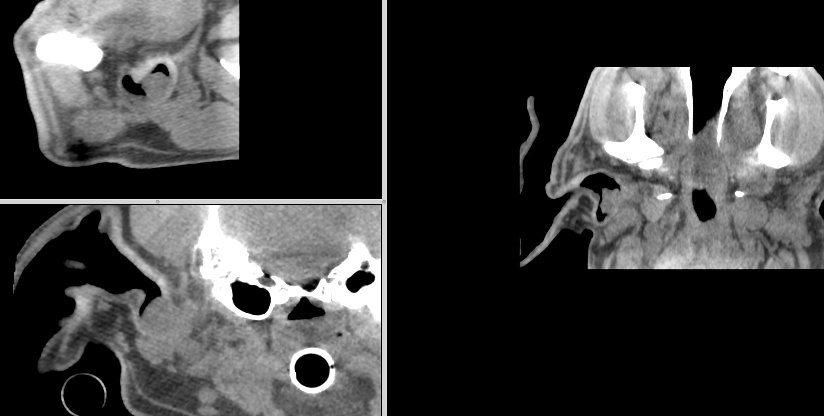
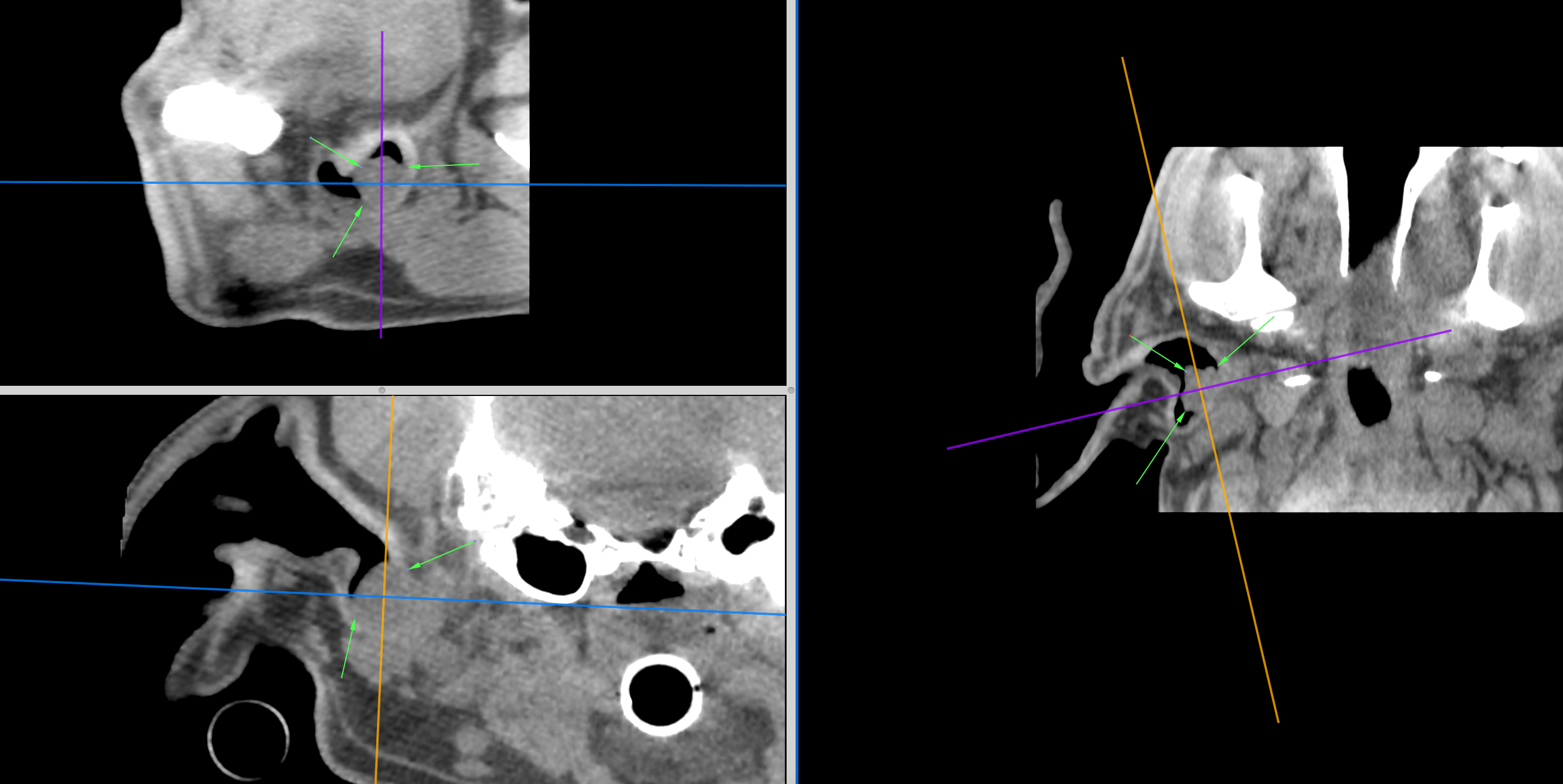
The patient presented for a mass in the right ear canal which was tentatively diagnosed as a cutaneous plasma cell tumor by FNA at the RDVM. There is a history of ear infections, currently being treated with Baytril. There is also a history of a cutaneous plasma cell tumor that was removed from the oral cavity in April 2015.
The patient presented for a mass in the right ear canal which was tentatively diagnosed as a cutaneous plasma cell tumor by FNA at the RDVM. There is a history of ear infections, currently being treated with Baytril. There is also a history of a cutaneous plasma cell tumor that was removed from the oral cavity in April 2015.
Physical Exam: BAR with a body condition score of 6/9. Eyes, ears, nose and throat WNL. A moderate amount of dental calculus was noted on the teeth. Gingiva were pink, CRT 2 seconds. The coat was healthy. No cardiac murmur or arrhythmias auscultated. BVS WNL. Abdominal palpation soft, non-painful. Neurologic system WNL. The gait was ambulatory x 4. Musculoskeletal system appeared to be NSF. Pain Scale (Colorado 0-4) – 0-1.