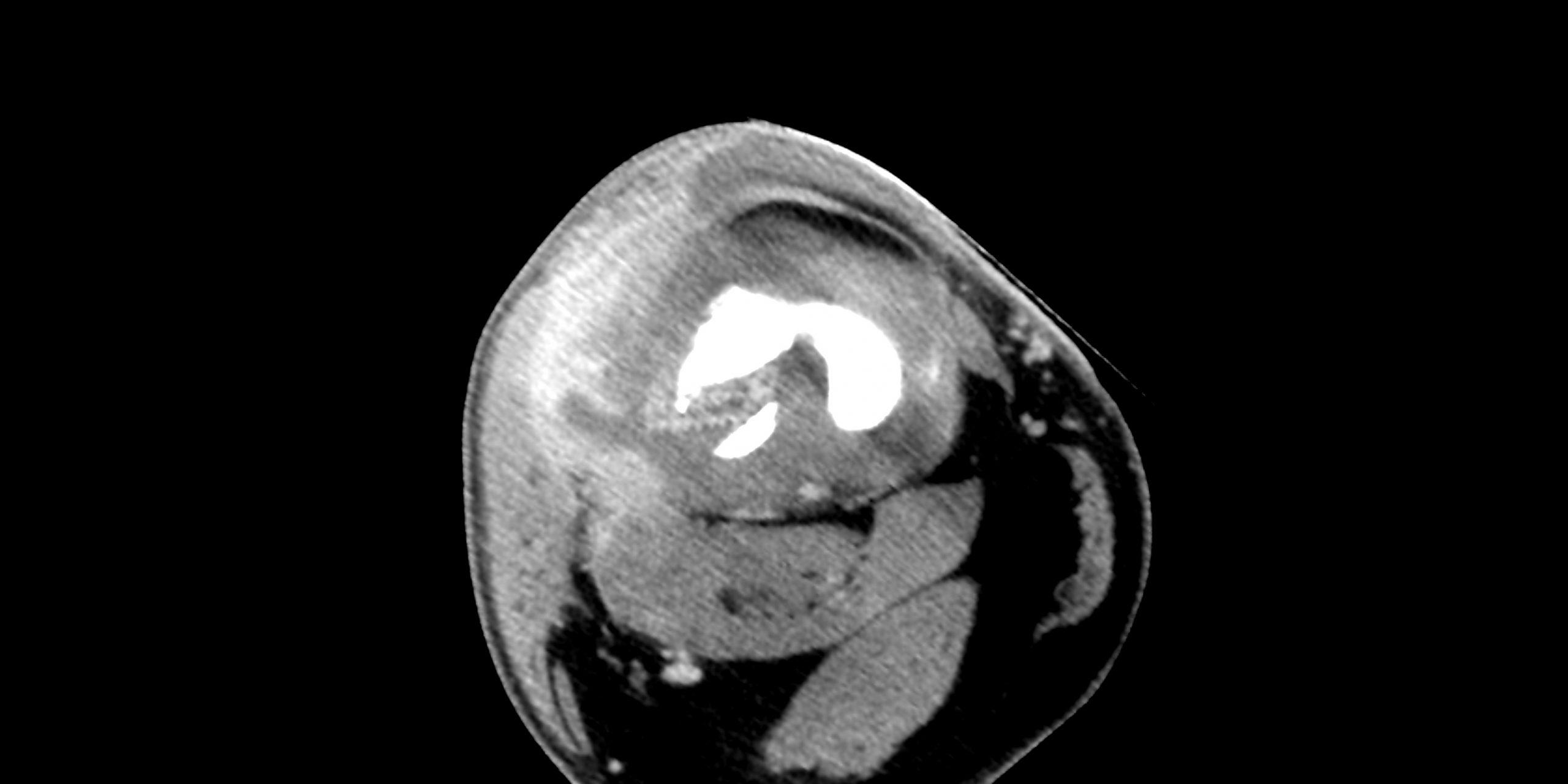
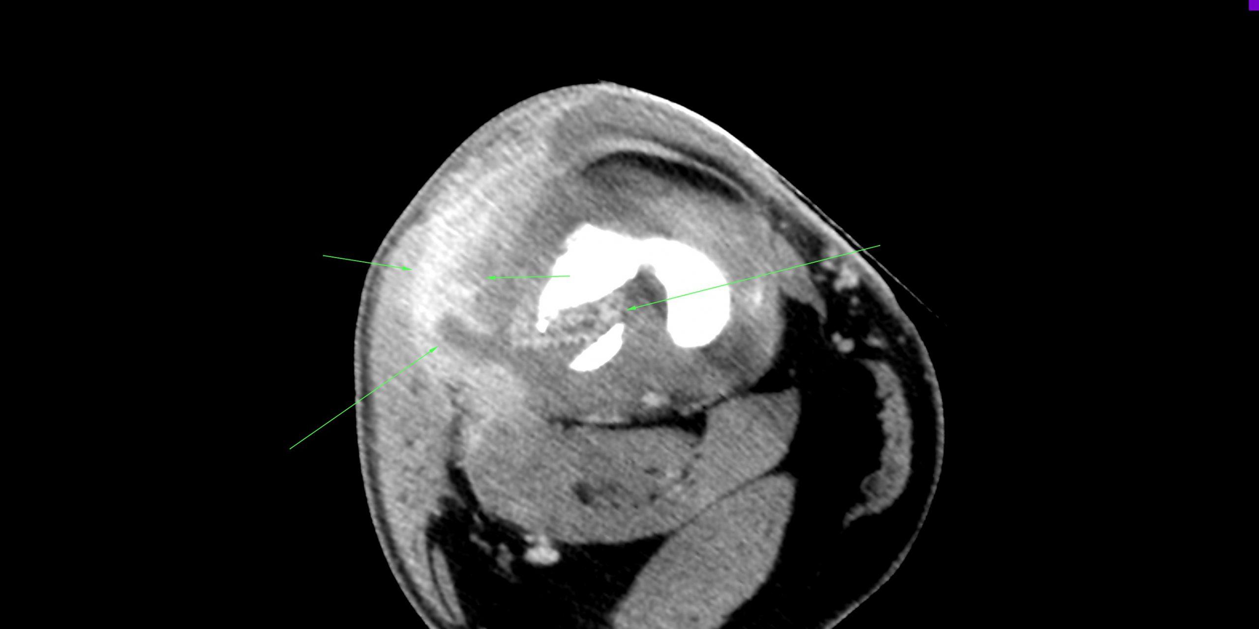
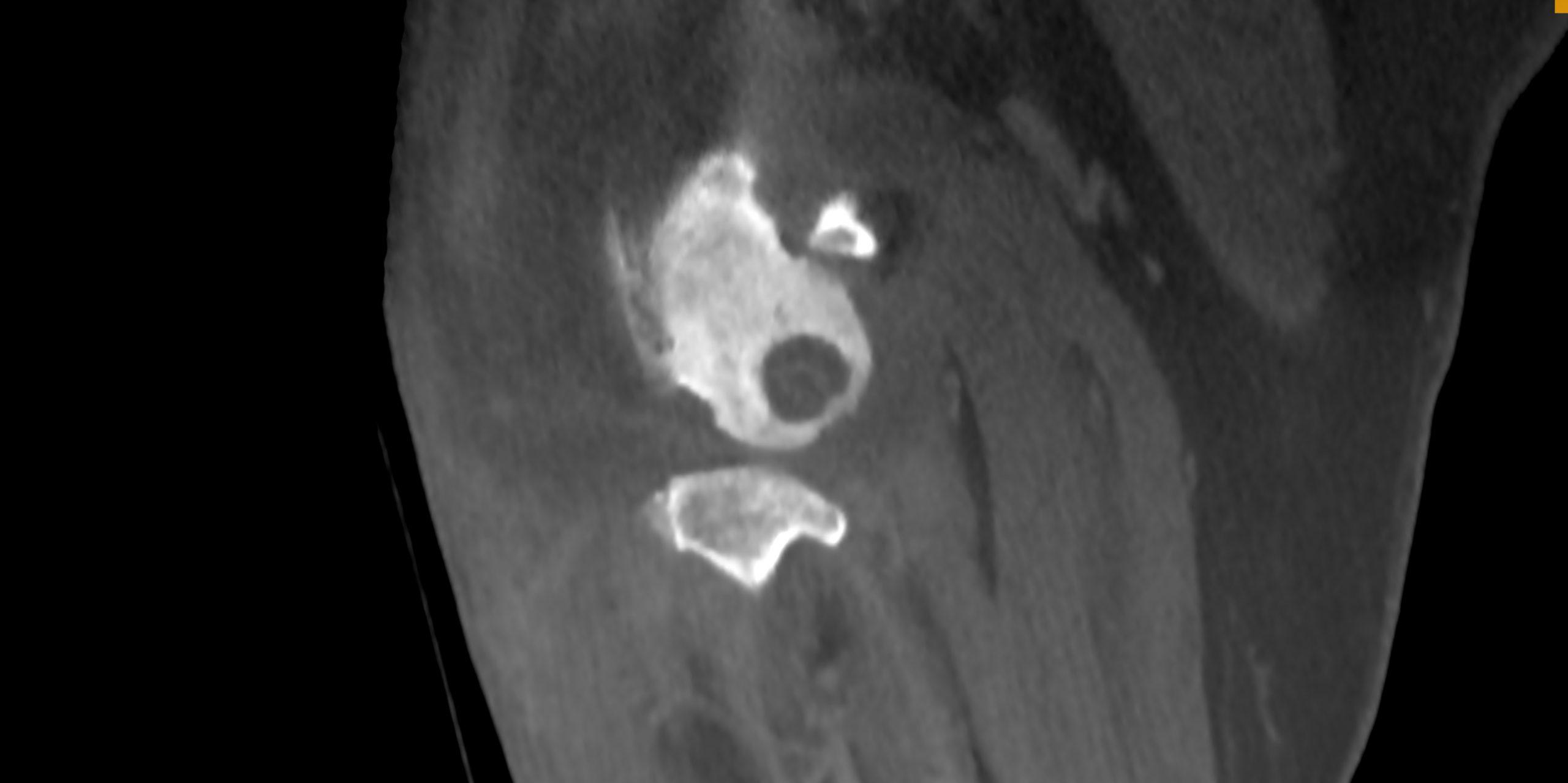
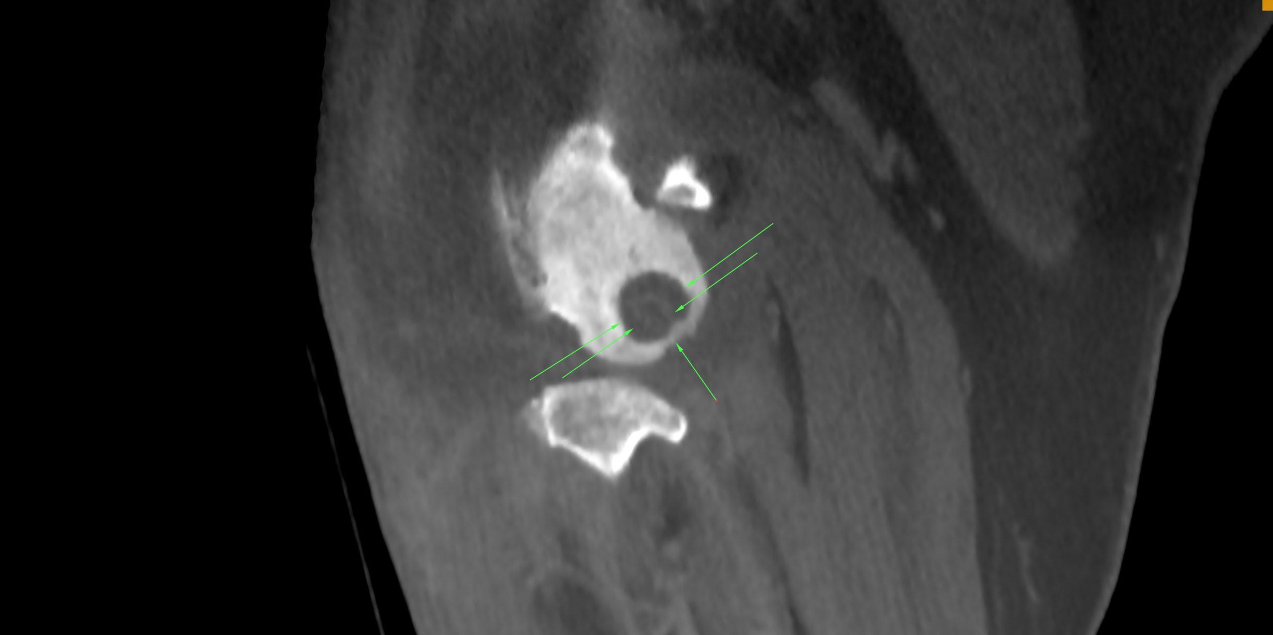
CT of the right stifle, plain and iodinated post contrast series: The scan showed a status after surgical correction for cranial cruciate ligament pathology using swivel lock anchor and fiber tape. There was significant widening and halo formation with reduction of bone mass and moderate sclerosis surrounding the femoral screw. The screw was off axis in respect to the formerly drilled canal. Mild widening surrounding the tibial bone anchor with mild sclerosis was noted. There was a moderate to severe amount of joint effusion within the femorotibial joint. Moderate joint capsule thickening and synovial proliferations were present. There was significant soft tissue swelling lateral to the stifle joint with irregularly increased contrast enhancement, emphasizing the region of the femoral condyle. Focal lateral capsular extension was noted lateral to the screw. Focal soft tissue thickening with rim enhancement pattern was seen in the intercondylar region of the stifle joint. A moderate amount of new bone formation which presented mildly unsharp in outline was seen at the periarticular margins. The lateral compartment of the femorotibial joint was decreased in height. The subchondral bone of the lateral femoral condyle showed significant sclerosis and a subchondral bone defect. The cranial cruciate ligament was not seen. Two popliteal lymph nodes were mildly enlarged in size but normal for shape, short-to-long-axis ratio and contrast uptake.