06_00259 Kimi S Urethral and bladder masses

Case Study

06_00259 Kimi S Urethral and bladder masses

A 7½-year-old SF Shiba Inu with a history of being on stillbestrol for urinary incontinence and recent UTI that had improved on antibiotics was presented for evaluation of pollakiuria. Abnormalities on urinalysis were hematuria, leukouria, and sheets of transitional epithelial cells.
 

Sonographic Differential Diagnosis

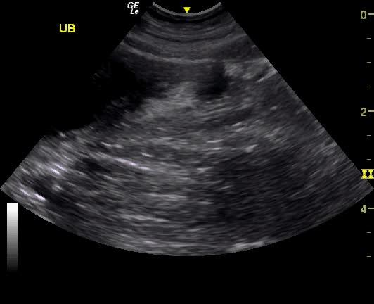
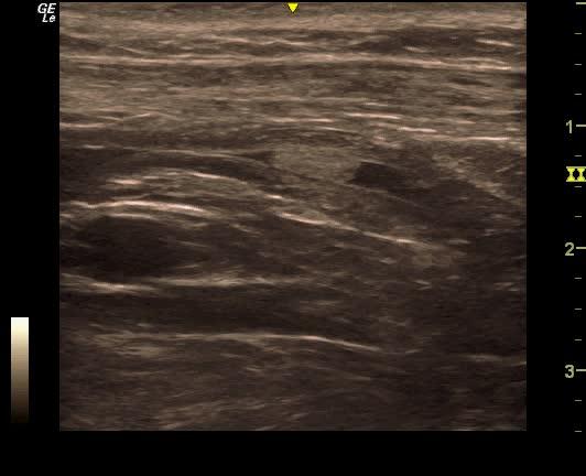
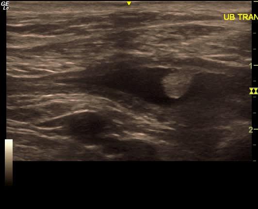
Non-resectable urethral and bladder wall mass, highly vascular on power Doppler. This is strongly suggestive for transitional cell carcinoma.

Image Interpretation

The urinary bladder in this patient presented a urethral mass that measured 0.5-0.7 cm with a separate midbody bladder mass that measured 3.18 x 2.02 cm. The urethral portion of the mass extended into the cystourethral junction.

DX

Urethral and bladder masses

Outcome

None

Clinical Differential Diagnosis

Bladder – neoplasia, urolith, chronic bacterial cystitis, granulomatous cystitis, polyploid cystitis
Urethra – neoplasia, granulomatous urethritis, liths

Sampling

None

Patient Information

Gender : Female, Spayed
Species : Canine
Type of Imaging : Ultrasound
Status : Complete

Clinical Signs

  • Pollakiuria

Images

vlcsnap-2015-06-28-17h40m59s62vlcsnap-2015-06-28-17h42m39s72vlcsnap-2015-06-28-17h45m30s14

Clinical Signs

  • Pollakiuria

Urinalysi

  • Blood Present
  • Epithelial Cells Present
  • WBCs Present