06_00256 Camden R Ectopic ureter

Case Study

06_00256 Camden R Ectopic ureter

A 1-year-old intact female Border collie that had been treated for a UTI was presented for polyuria and urinary incontinence. Physical examination was normal. The only abnormalities on urinalysis were the presence cocci bacteria and struvite crystals.


Sonographic Differential Diagnosis

Left ectopic ureter, appears to be entering into the urethra and appears extramural. Surgical consultation is recommended or non-invasive approach with laser ablation performed at Animal Medical Center in New York with Dr. Berent.

Image Interpretation

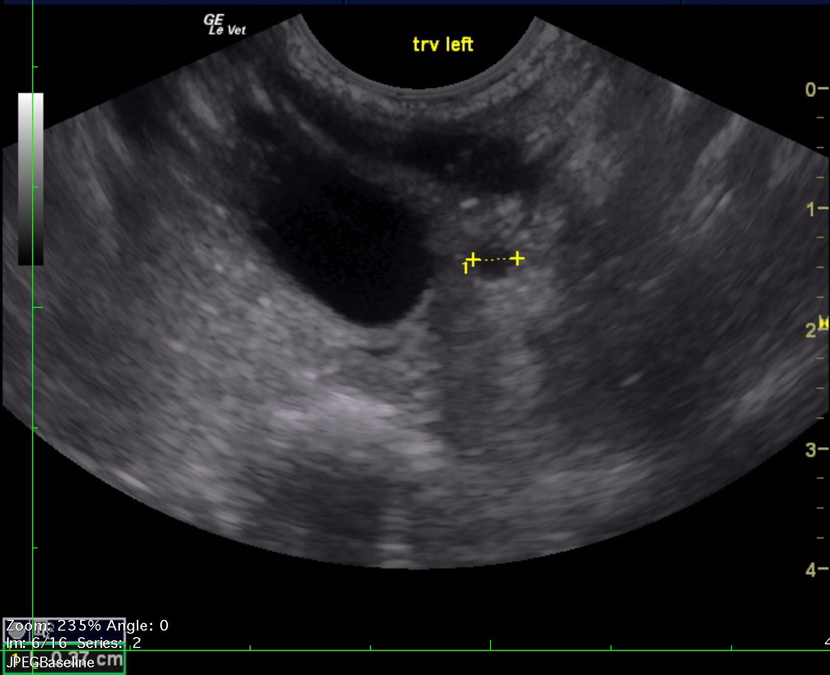
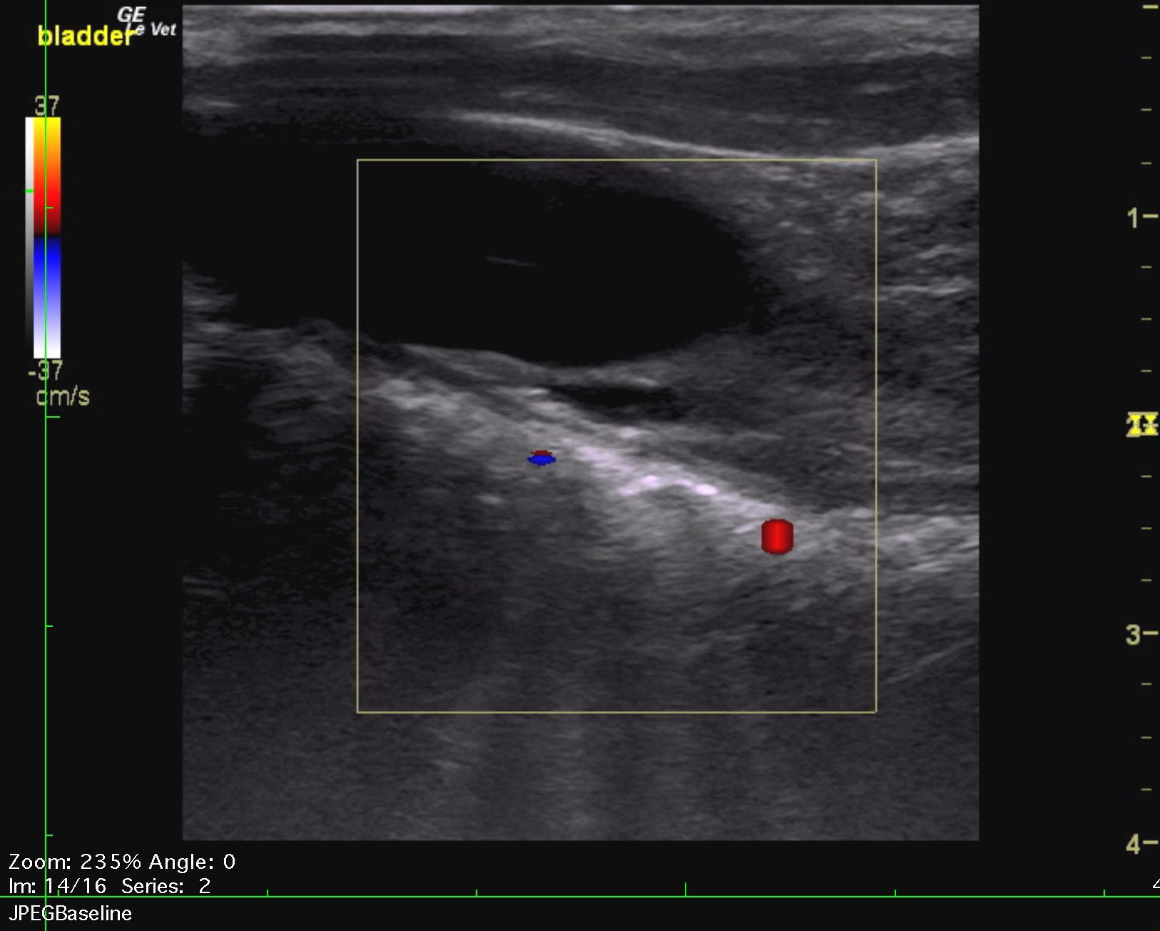
The urinary bladder revealed an ectopic ureter that appeared to be entering into the pelvic urethra and measured 0.5 cm at the level of the trigone. The remainder of the bladder was unremarkable. The uterus was uniform and measured 0.76 cm.
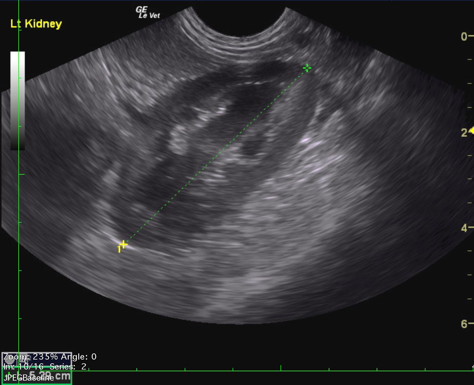
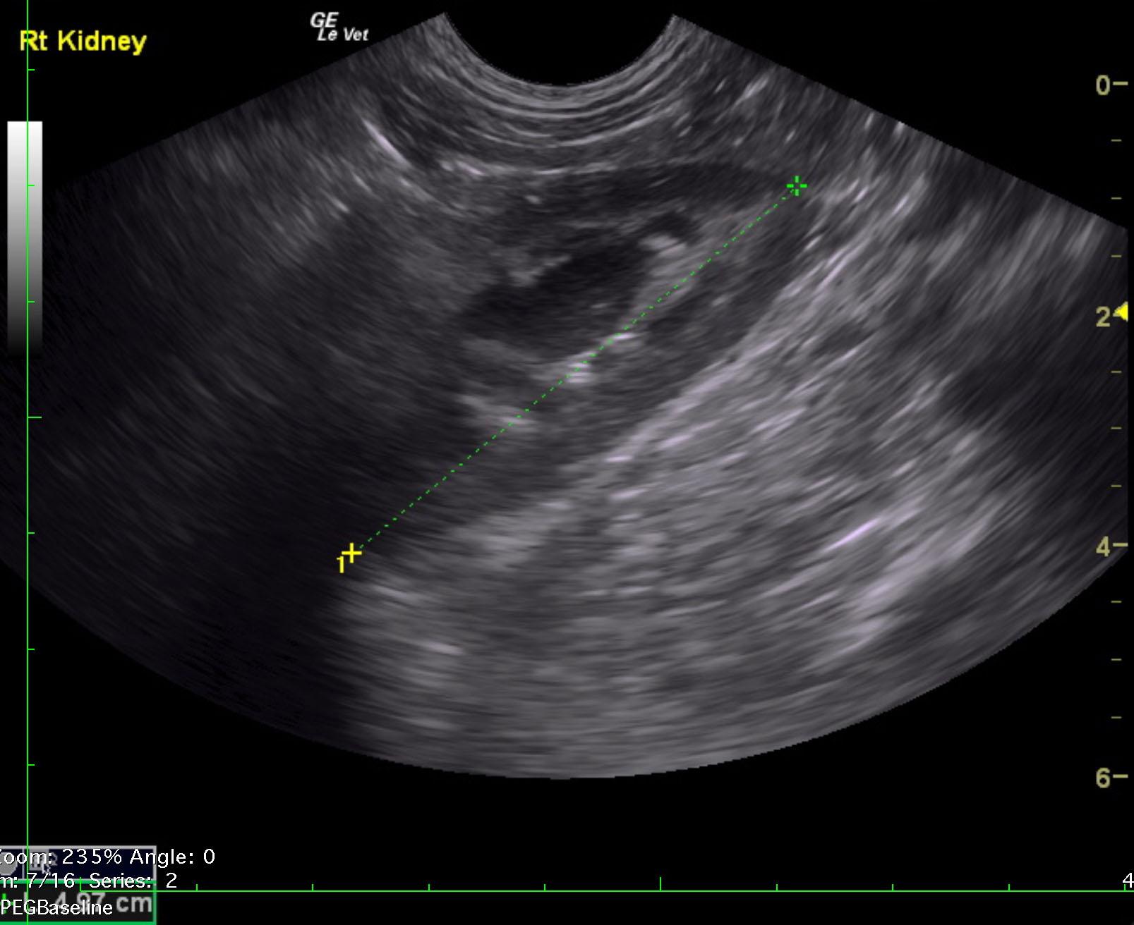
The kidneys revealed normal size and structure, corticomedullary definition and ratio (cortex 1/3 of medulla). The cortices presented largely uniform texture with normal echogenic relationship to liver and spleen. Medullary echogenicity differed distinctly from that of the cortex and no evidence or dilation could be seen. The capsules were acceptably uniform without dramatic irregularities. The left kidney measured 5.3 cm and the right kidney measured 4.97 cm. A slight amount of free fluid was noted.

DX

Unilateral ectopic ureter

Outcome

None

Clinical Differential Diagnosis

Ectopic ureter, chronic bacterial cystitis, uroliths

Sampling

None

Patient Information

Gender : Female, Intact
Species : Canine
Type of Imaging : Ultrasound
Status : Complete

Clinical Signs

  • Incontinence
  • Polyuria

Images

rimmer_camden_left_ectopic_ureterrimmer_camden_left_ectopic_ureter_cfrimmer_camden_left_ectopic_ureter_lkrimmer_camden_left_ectopic_ureter_rk

Clinical Signs

  • Incontinence
  • Polyuria

Urinalysi

  • Bacteria Present
  • Struvite crystals
Skip to content