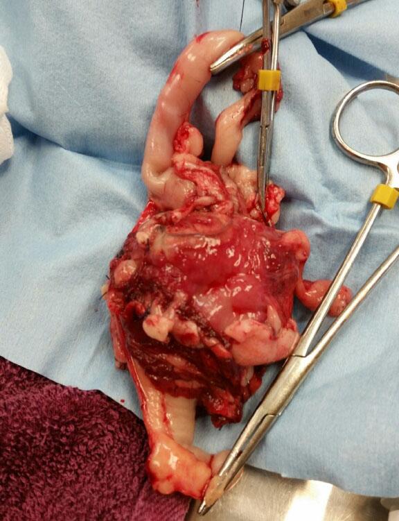
04_00458 Kirby F Jejunal mass

Case Study

04_00458 Kirby F Jejunal mass

A 14-year-old SF DSH was presented for evaluation of pyrexia and weight loss. Urinalysis showed an SG of 1.023. Abnormalities on CBC and serum biochemistry were severe anemia, elevated BUN (12), total bilirubin, amylase, and triglycerides. Coagulation panel showed severely elevated APTT and mildly elevated PT.
 

Sonographic Differential Diagnosis

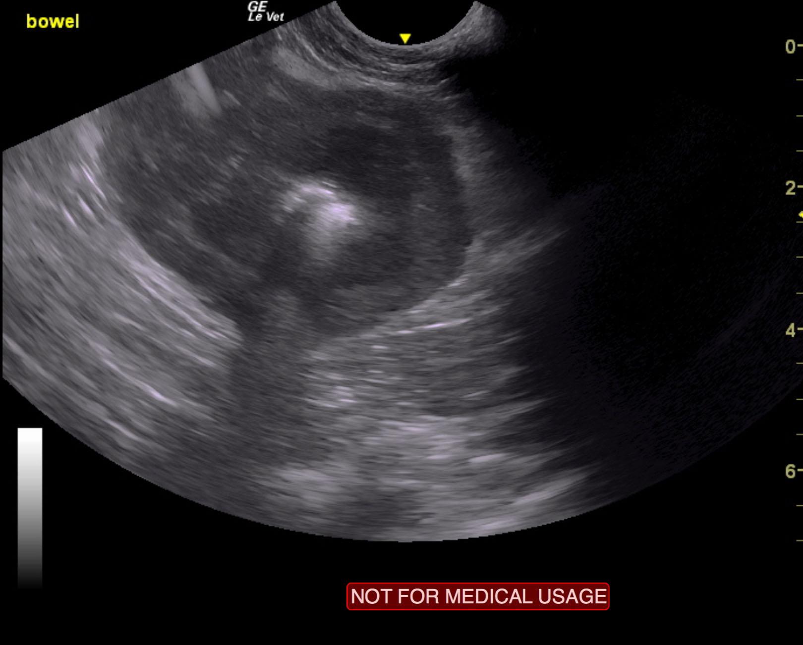
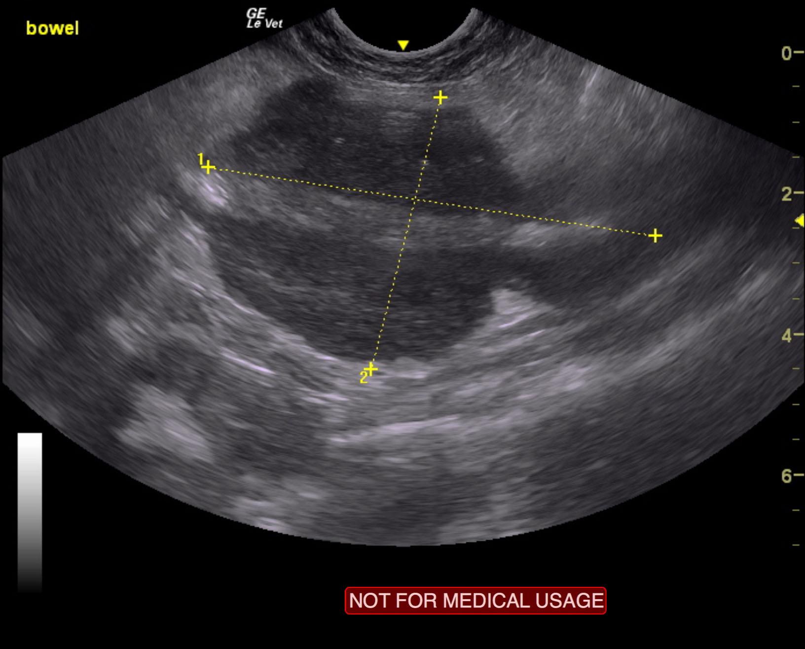
Primary intestinal mass with regional lymphadenopathy and reactive fat. Variable small intestinal thickening elsewhere. Epigastric lymphadenopathy.

Image Interpretation

The gastrointestinal tract revealed an irregular bowel mass that was deriving from the wall. Deviation of the lumen was noted. This measured 5.47 x 4.2 cm with annular strictured contour. Hyperechoic, ill defined surrounding mesentery measuring 6.4 x 4.0 cm. Variable, other portions of the intestine appeared to be thickened with minor loss of mural detail. Hypoechoic, irregular epigastric lymph node was noted.

DX

Jejunal mass

Outcome

None

Clinical Differential Diagnosis

Hemolytic anemia, FIV/FeLV, FIP
Neoplasia – lymphoma, mast cell tumor
Liver – chronic hepatopathy, cirrhosis, neoplasia, granulomatous disease, cholangio-hepatitis complex

Sampling

None

UA Specific Gravity Range

1.023

Patient Information

Gender : Female, Spayed
Species : Feline
Type of Imaging : Ultrasound
Status : Complete

Clinical Signs

  • Fever
  • Weight loss

Images

kirby_fletcher_intestinal_mass_lsa_felinekirby_fletcher_intestinal_mass_lsa_feline_2img_4261

Blood Chemistry

  • Amylase, High
  • BUN high
  • Hypertriglyceridemia
  • Total Bilirubin, High

CBC

  • RBC, Low

Clinical Signs

  • Fever
  • Weight loss

Special Testing

  • PT Prolonged
  • PTT Prolonged