A 4-year-old SF Norwich Terrier dog was presented for one-week duration of intermittent anorexia and constipation. Abnormalities on blood work included mild anemia, elevated ALT activity, and low BUN. The dog was also Lyme positive.
A 4-year-old SF Norwich Terrier dog was presented for one-week duration of intermittent anorexia and constipation. Abnormalities on blood work included mild anemia, elevated ALT activity, and low BUN. The dog was also Lyme positive.
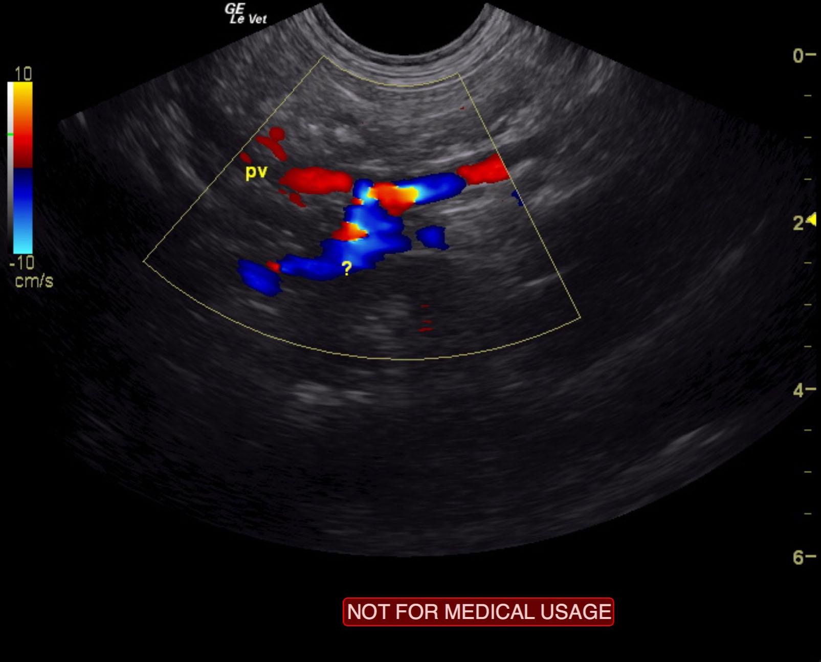
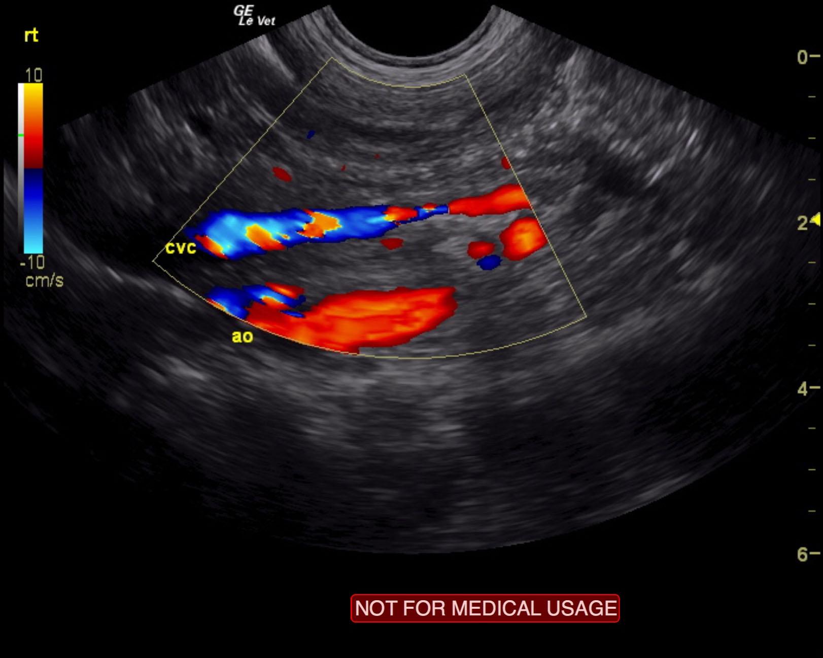
Extrahepatic portosystemic shunt, likely splenoazygos. Microhepatica.
The liver was significantly subnormal in size at 2.1 cm in short axis. A splenazygous shunt was noted in this patient and measured 0.85 cm in width and approximately 1.8 cm in length prior to entering dorsally into the vena cava. The pre-shunt portal vein measured 0.5 cm. Vena cava after the shunt measured 0.5 cm, and the aorta was 0.6 cm. Residual portal vein 0.3 cm. It appears that the shunt exits the splenic entry to the portal vein and creates a “double aorta” appearance cranial to the diaphragm, which is most consistent with a splenoazygos shunt. Hepatic parenchyma was hypovascular and relatively uniform with slightly increased portal markings. Concurrent inflammatory hepatopathy is likely along with the extrahepatic portosystemic shunt. The gallbladder was unremarkable.
None
Hepatic disease: resolving acute hepatitis (viral, bacterial), chronic-active hepatitis, porto-caval shunt, primary portal vein hypoplasia, cirrhosis (congenital, acquired).
None