04-00456 Louis M IBD —NEED SL—-

Case Study

04-00456 Louis M IBD —NEED SL—-

A 13-year-old NM DSH was presented for evaluation of chronic vomiting and weight loss. Abnormalities on blood work were mild hypoalbuminemia, hyperglobulinemia, and mild hypocalcememia.

A 13-year-old NM DSH was presented for evaluation of chronic vomiting and weight loss. Abnormalities on blood work were mild hypoalbuminemia, hyperglobulinemia, and mild hypocalcememia.

Sonographic Differential Diagnosis

IBD GI pattern. Fine needle aspirates of the jejunal lymph nodes were performed without complication. PARR or PCR analysis may be appropriate on the cytology of those lymph nodes to ensure that underlying lymphoma is not an issue. Clinical trial of Zithromax, Prednisolone, and B12 injections along with a hypoallergenic diet. Full thickness GI biopsies may be necessary for definitive diagnosis.

Image Interpretation

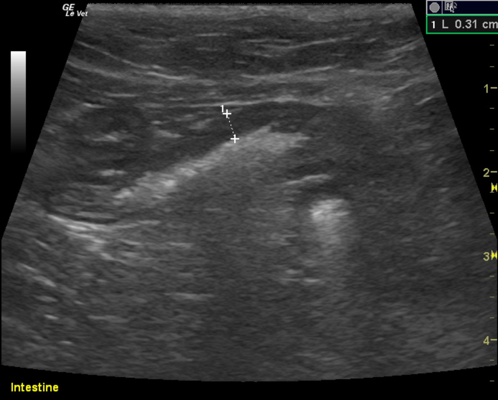
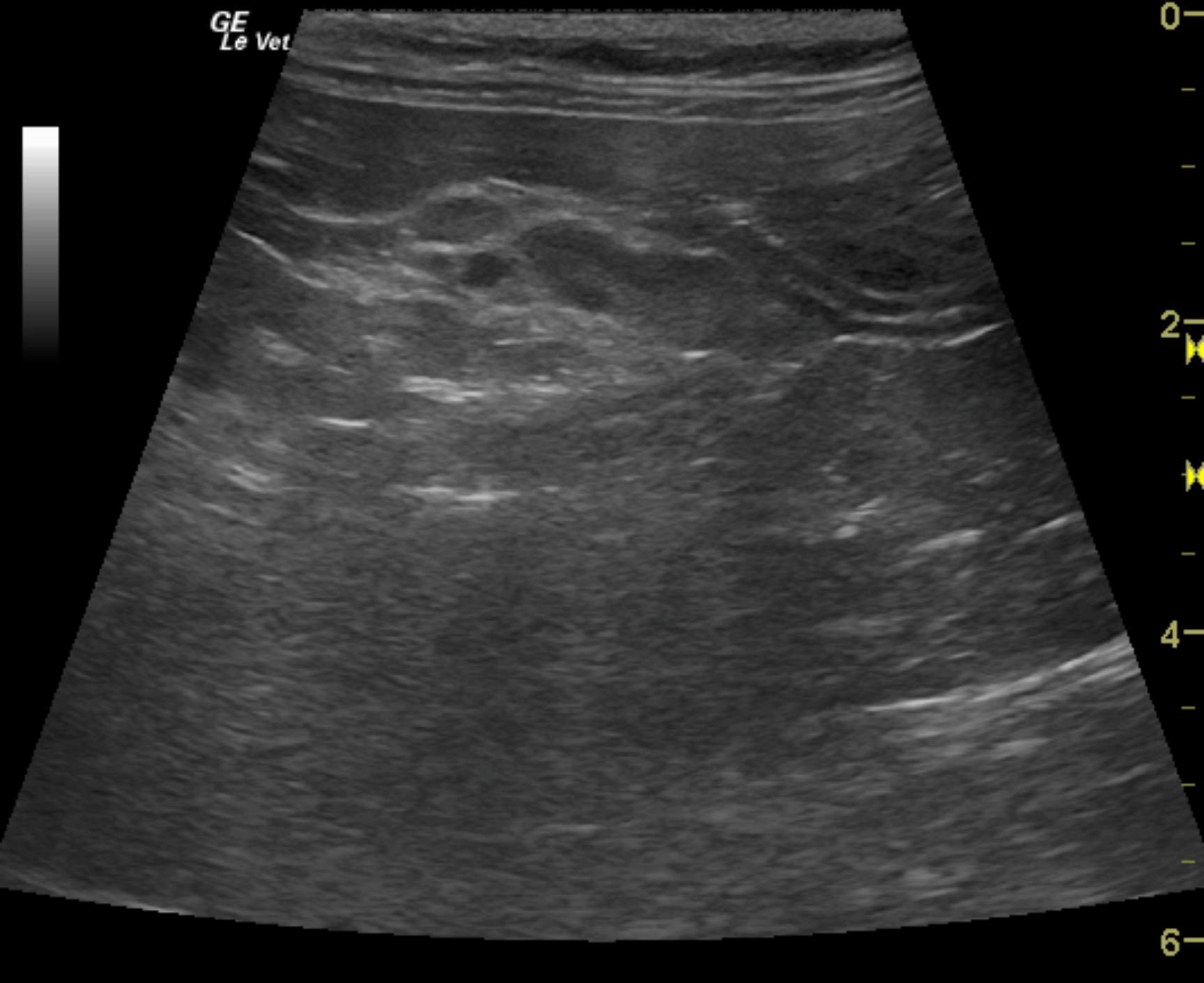
The gastrointestinal presentation revealed mild uniform prominence of the gastric mucosa as well as areas of “ropey” small intestinal wall. The intestinal mucosa was slightly irregular, thickened and hyperechoic suggestive of low grade, chronic inflammation. The muscularis and variable portions of the small intestine revealed a 1:1 muscularis mucosa ratio 0.31 cm thickness of the small intestine. This is an idiopathic finding, which may be related to variable forms of inflammatory bowel and intestinal neoplasia. However, no loss of detail was noted. Segmental hypertrophy of the intestinal wall was noted in variable areas of the small intestine. Ileocecal lymph node slightly enlarged at 2.04 x 0.79 cm yet uniform consistent with reactive lymph node. Some reactive hyperechoic ill-defined fat was noted associated with the mesenteric lymph nodes and portions of the small intestines most consistent with a flare up of inflammatory bowel. Mild potential for emerging lymphoma, mast cell disease, dry form FIP.

DX

IBD

Outcome

None

Clinical Differential Diagnosis

GIT – neoplasia, IBD, dietary hypersensitivity, ulceration, foreign body, granulomatous enteritis
Pancreas – chronic pancreatitis, neoplasia, abscessation

Sampling

Jejunal LN

Patient Information

Gender : Male, Neutered
Species : Feline
Type of Imaging : Ultrasound

Clinical Signs

  • Vomiting
  • Weight loss

Images

mackinnon_louis_ibd_panc_muscularis_hypertrophy_20150525081014

Blood Chemistry

  • Albumin, Low
  • Calcium, Low
  • Globulin, Low

Clinical Signs

  • Vomiting
  • Weight loss