An 11-year-old NM DSH was presented for evaluation of weight loss and intermittent vomiting.
An 11-year-old NM DSH was presented for evaluation of weight loss and intermittent vomiting.
An 11-year-old NM DSH was presented for evaluation of weight loss and intermittent vomiting.
An 11-year-old NM DSH was presented for evaluation of weight loss and intermittent vomiting.
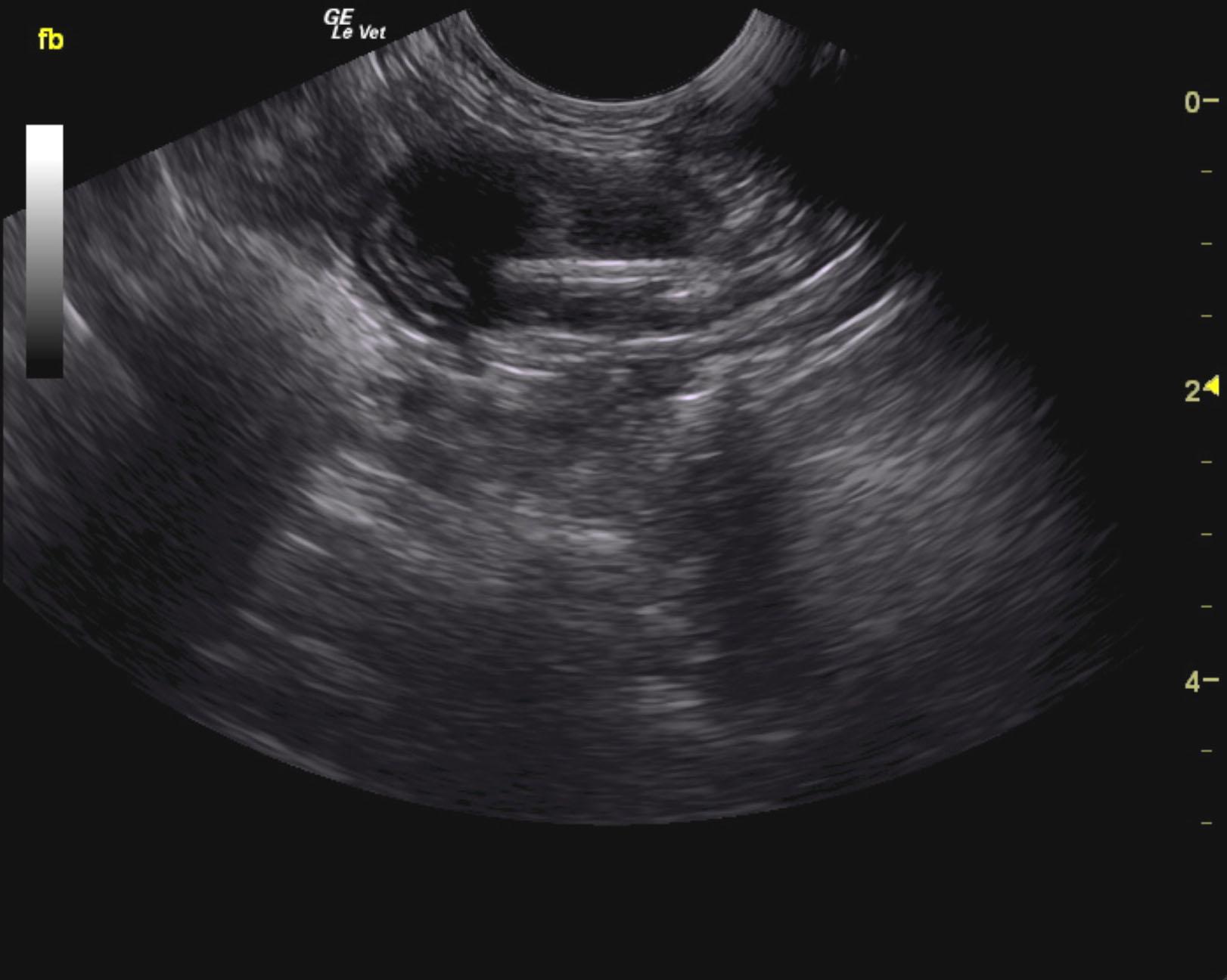
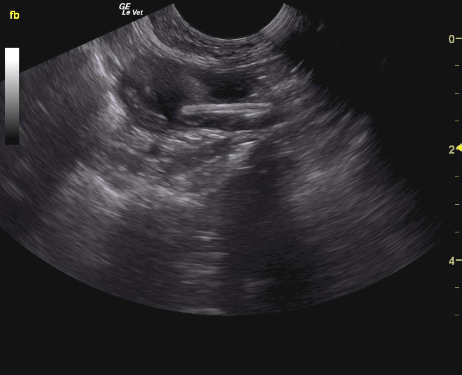
Diffuse IBD gastrointestinal pattern with focal intestinal wall thickening and foreign structure. There is a strong potential for underlying intestinal mural neoplasia along with foreign matter. Intraoperative ultrasound and resection and anastamosis would be ideal. There was a minor obstructive pattern. If the surgeon is to perform resection and anastamosis without ultrasound-guidance then at least 5 cm of small bowel should be removed proximal to and distal from the focal mural thickening within the bowel, which measures approximately 1.5 cm in width. This extends for approximately 2.0 cm. Slight hyperechoic focus was noted within the lumen and would suggest foreign matter. However, it is likely that the foreign matter is not the primary issue. Underlying histopathology is very important in this case as underlying lymphoma or focal carcinoma is possible or complicated inflammatory bowel.
The gastrointestinal tract revealed a diffuse muscularis thickening with a 1:1 ratio with increased submucosal echogenicity and a focal, 1.5 cm linear type foreign body noted embedded within the thickened portion of small intestine. This measured approximately 1.0 cm. Resection of this portion of bowel along with removal of the focal foreign structure is recommended. Minor focal accumulation was noted within the small intestine. It appears that the portion of bowel is corrugated around the foreign body.
None
GIT – IBD, dietary hypersensitivity, neoplasia, foreign body, ulceration, granulomatous enteritis, helminths
Hyperthyroidism
Pancreas – chronic pancreatitis, neoplasia
None