03_00424 Geronimo Z EHBDO

Case Study

03_00424 Geronimo Z EHBDO

An 8 year-old, MN, DSH feline was presented for abdominal ultrasound for weight loss, anorexia and icterus. With blood work demonstrating a bilirubin 5.6, elevated liver enzymes, and elevated coags. 
An 8 year-old, MN, DSH feline was presented for abdominal ultrasound for weight loss, anorexia and icterus. With blood work demonstrating a bilirubin 5.6, elevated liver enzymes, and elevated coags. 
Sonographic Differential Diagnosis

The appearance of the liver could certainly be due to cholangitis, which can either be due to lymphocytic plasmacytic inflammation or suppurative inflammation. Lymphoma is another possibility and cannot be excluded. The appearance of the liver was not consistent with hepatic lipidosis. Obstructive lesion at the duodenal papilla resulting in bile duct obstruction and increased bilirubin. This could be due to poorly calcified stone or a soft tissue structure which I think would be less likely based on appearance. Prominent pancreas can be due to concurrent pancreatitis but can also be within normal limits. A surgical exploratory is strongly advised given the presence of the structure within the bile duct and the concern of obstructed bile duct. A liver biopsy and a culture of the bile should be obtained at the time of surgery. Continue supportive care with fluids, antibiotics, antiemedics and pepcid and pain medication as needed.

Image Interpretation

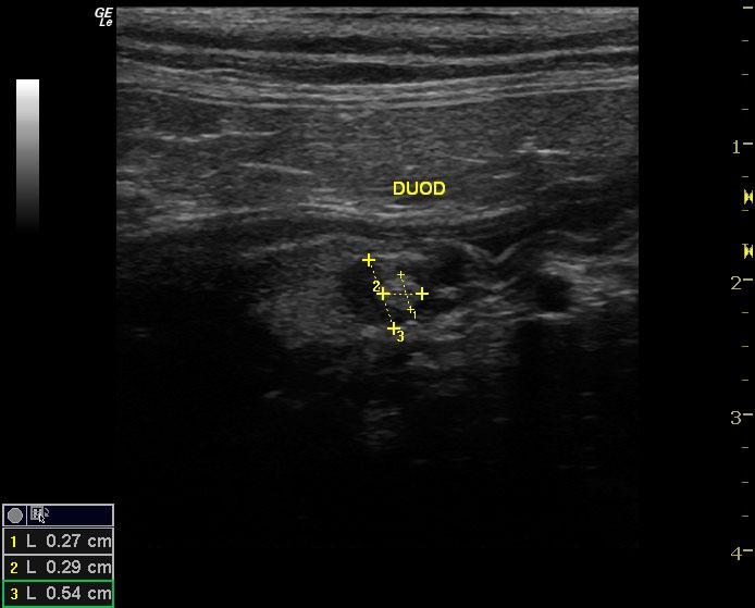
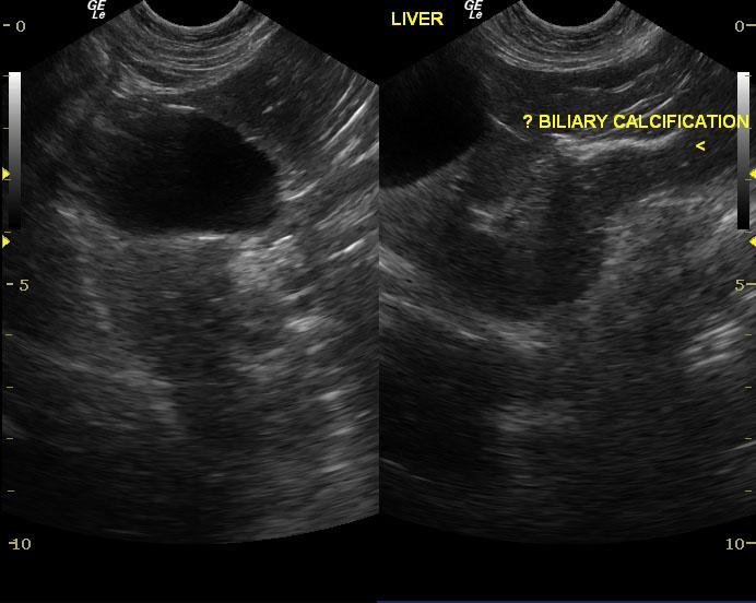
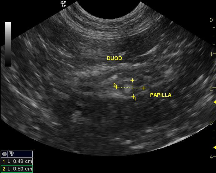
The liver was homogenous and appeared mildly decreased in echogenicity. There were linear segments of biliary mineralization. There was no evidence of any masses or nodules within the liver. The contours of the liver were normal. The gallbladder was normal and there was no evidence of any masses or choleliths or sludge within the gallbladder. The gallbladder was distended. The bile duct was dilated especially at the duodenal papilla measuring up to 0.5cm in diameter at that point. There was a hyperechoic round structure located within the bile duct at the duodenal papilla and appeared to be obstructive and measured 0.27 x 0.29cm. This hyperechoic structure did not shadow. The pancreas was prominent but did not appear overtly inflamed.

DX

EHBDO

Outcome

None

Sampling

None

Patient Information

Gender : Male, Neutered
Species : Feline
Type of Imaging : Ultrasound
Status : Complete

Clinical Signs

  • Anorexia
  • Icterus
  • Weight loss

Images

010911070824500109110708242301091107082432

Blood Chemistry

  • Elevated Liver Enzymes
  • Total Bilirubin, High

Clinical Signs

  • Anorexia
  • Icterus
  • Weight loss

Special Testing

  • PT Prolonged
  • PTT Prolonged