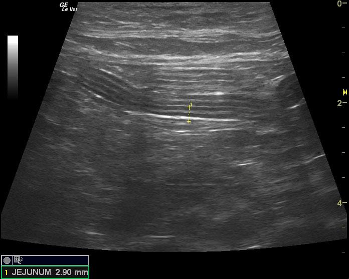
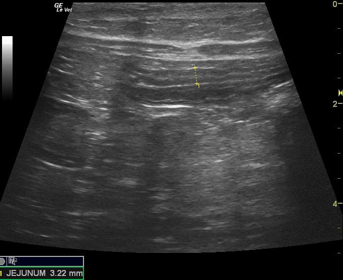
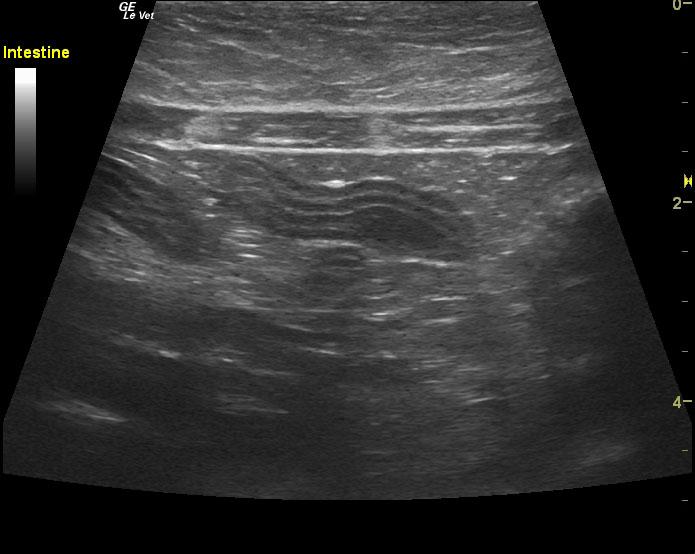
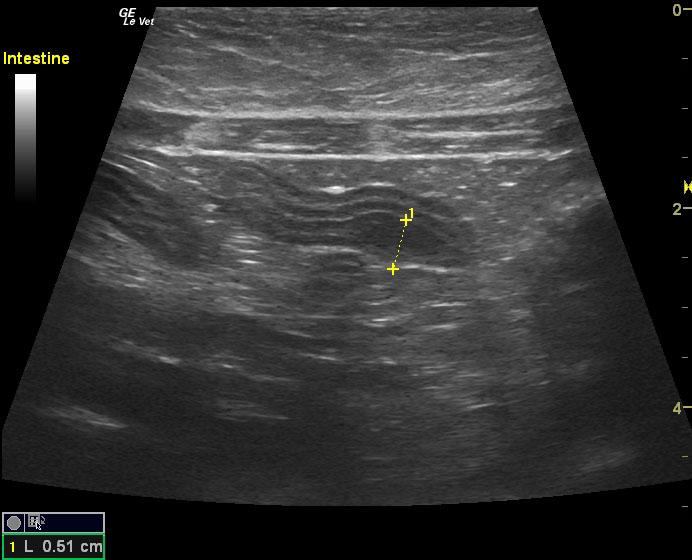
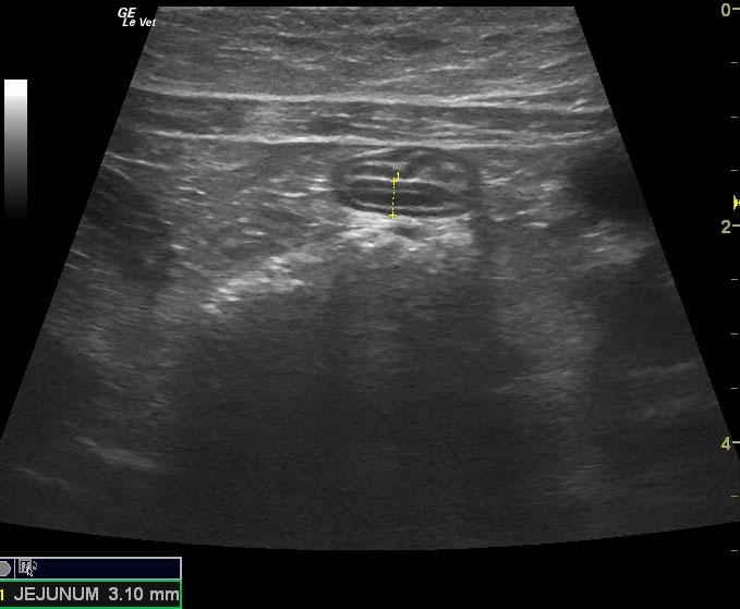
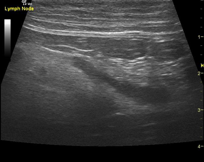
A 10-year-old NM DSH was presented for evaluation of intermittent vomiting and weight loss. On abdominal radiographs a slight accordion pattern was present.
A 10-year-old NM DSH was presented for evaluation of intermittent vomiting and weight loss. On abdominal radiographs a slight accordion pattern was present.