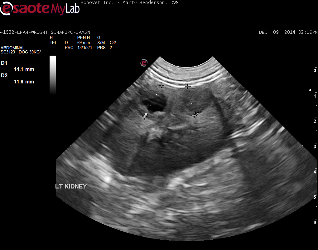
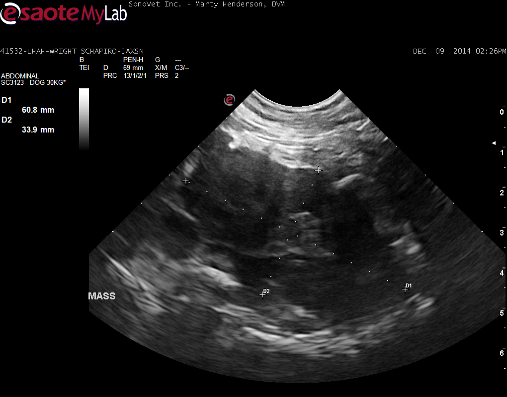
A 7-year-old MN Shepherd Mix was presented for evaluation of tenesmus with no other obvious signs. On rectal palpation a mass on the ventral aspect of the lumbar spine that starts approximately 0.5 cm into the pelvic canal and extends cranial for about an inch
A 7-year-old MN Shepherd Mix was presented for evaluation of tenesmus with no other obvious signs. On rectal palpation a mass on the ventral aspect of the lumbar spine that starts approximately 0.5 cm into the pelvic canal and extends cranial for about an inch