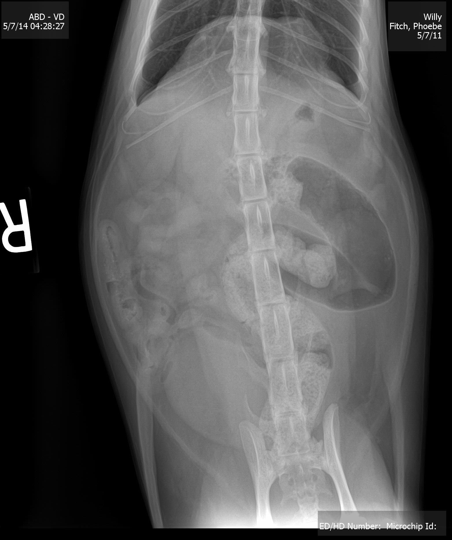
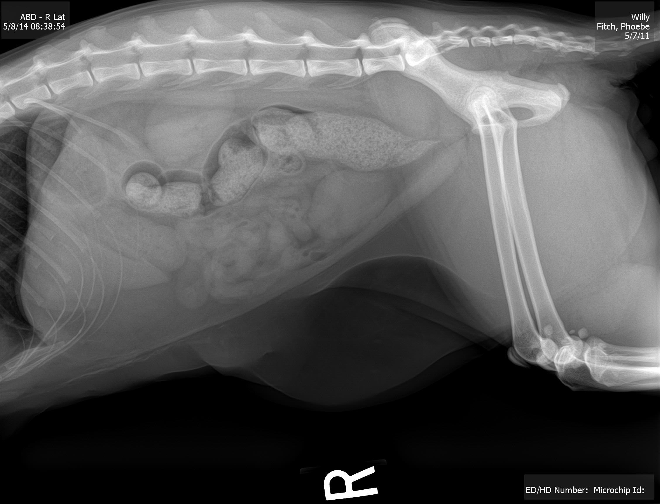
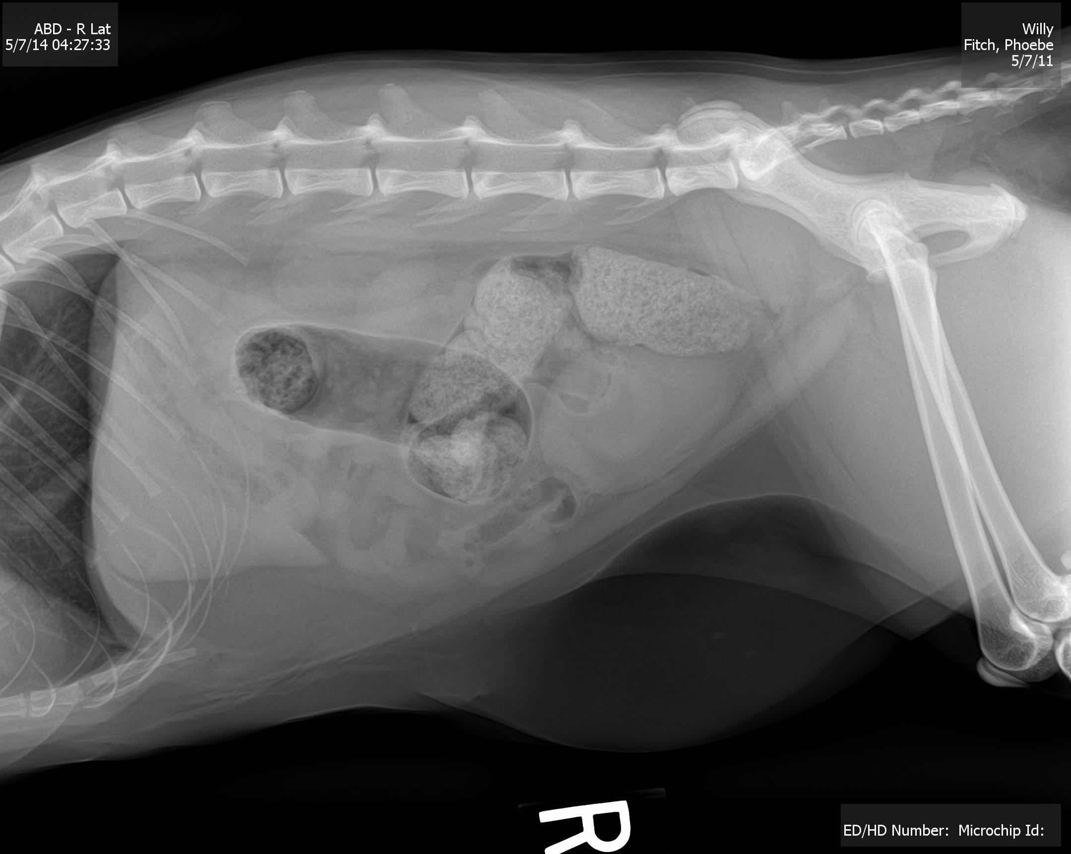
A 3-year-old NM DSH was presented for evaluation of vomiting and diarrhea. Radiographs showed constipation and narrowing of caudal colon by a dorsal mass effect compressing the colon. On rectal exam a large smooth firm mass was present.
A 3-year-old NM DSH was presented for evaluation of vomiting and diarrhea. Radiographs showed constipation and narrowing of caudal colon by a dorsal mass effect compressing the colon. On rectal exam a large smooth firm mass was present.
Descending colonic or pelvic lymph node mass. This appears isolated and potentially resectable. There is a potential for granuloma, carcinoma or lymphoma. Ultrasound-guided FNA are recommended for definition followed by potential surgical, oncological or radiation treatment. Resection may be possible; however, splitting the pelvic bone would be necessary.
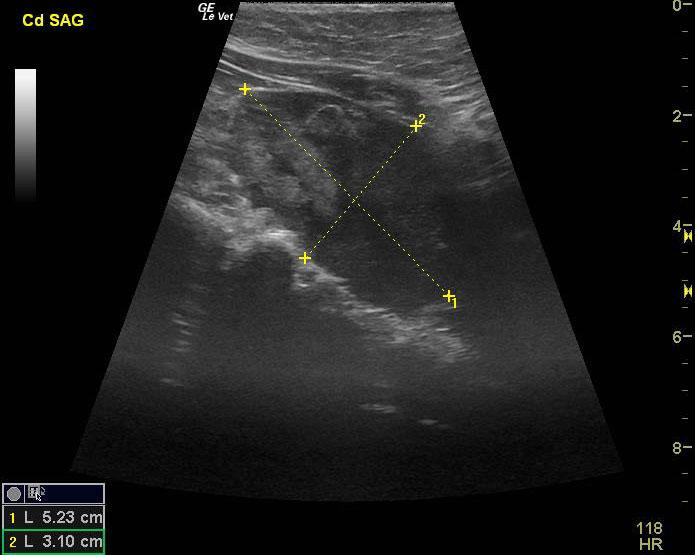
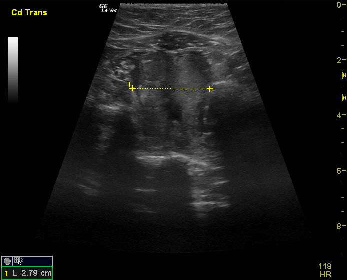
An irregular pelvic mass was noted and impinged the descending colon. The mass may be deriving from the descending colonic wall. The mass was echogenic and moderately complex measuring 5.23 x 3.1 cm. This mass appeared to be isolated.
None
Neoplasia, granulomatous colitis (FIP, fungal, foreign body), abscess
None