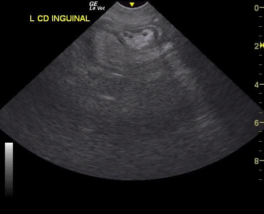
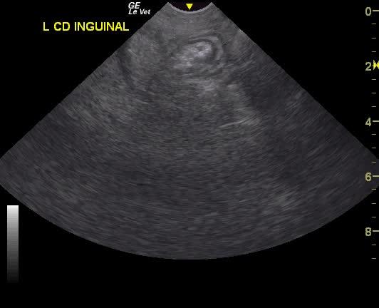
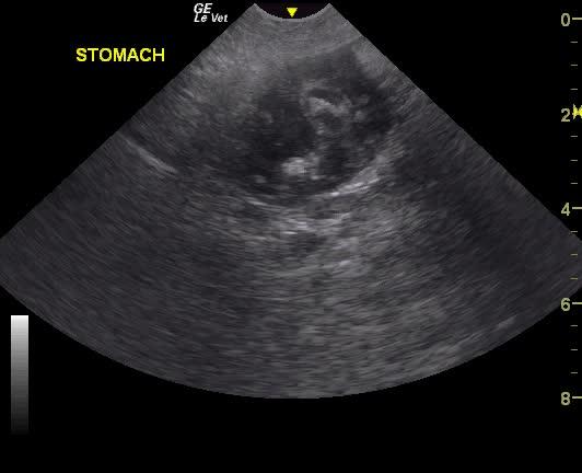
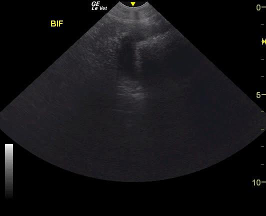
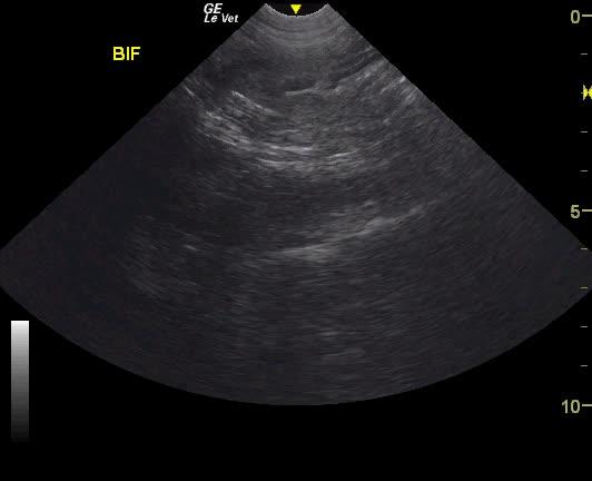
A NM Labrador with a history of foreign body ingestion was presented for intermittent diarrhea that had initially responded to bland diet, metronidazole and aminopentamide. More recently there had vomiting and he appeared uncomfortable. On physical examination, a palpable mass in the cranial abdomen was present. CBC was within normal limits and serum biochemistry showed elevated phosphate low BUN.
A NM Labrador with a history of foreign body ingestion was presented for intermittent diarrhea that had initially responded to bland diet, metronidazole and aminopentamide. More recently there had vomiting and he appeared uncomfortable. On physical examination, a palpable mass in the cranial abdomen was present. CBC was within normal limits and serum biochemistry showed elevated phosphate low BUN.