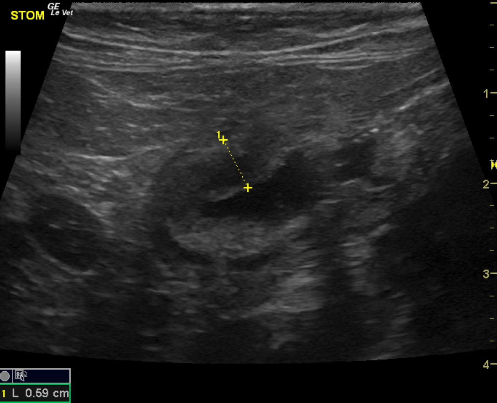
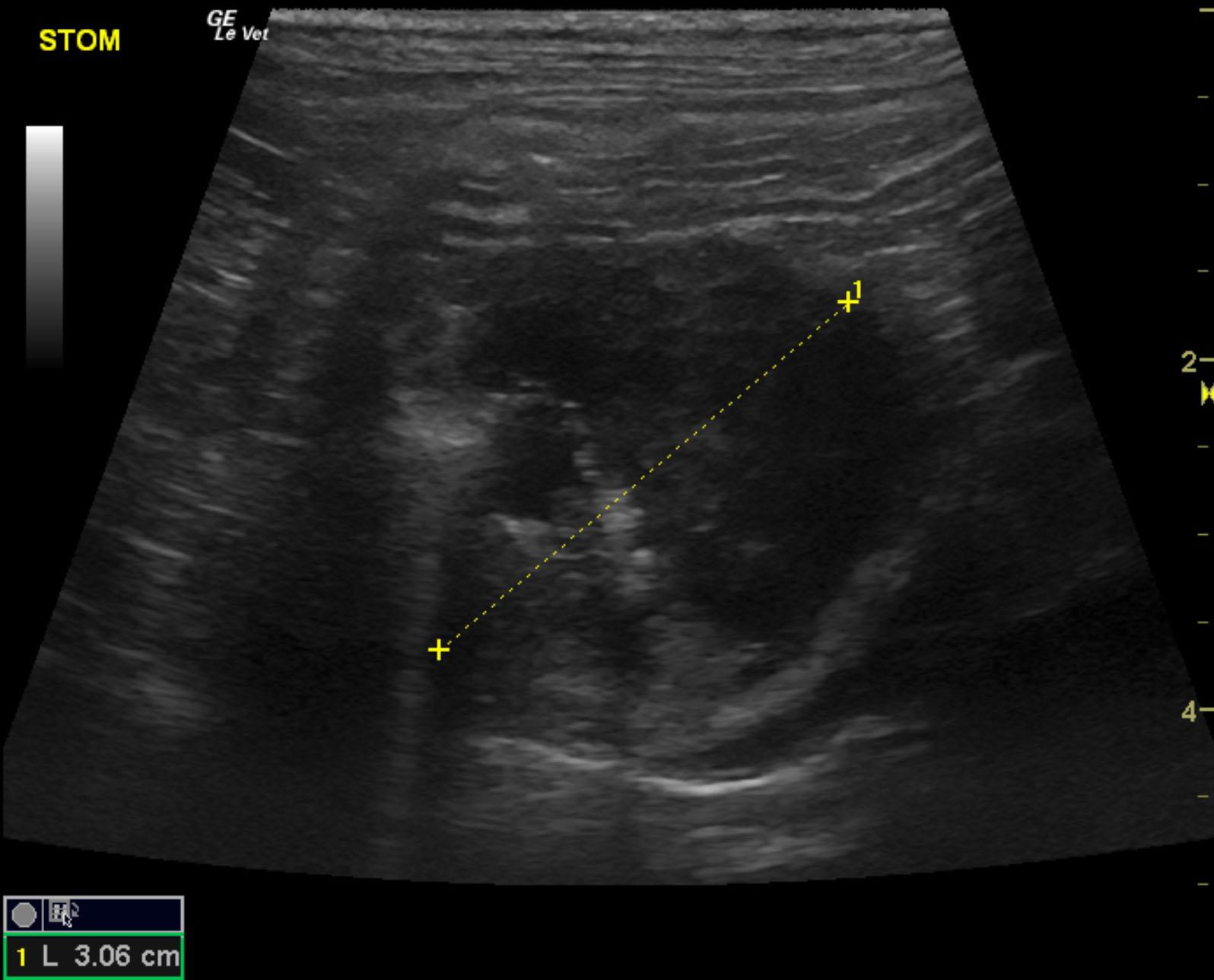
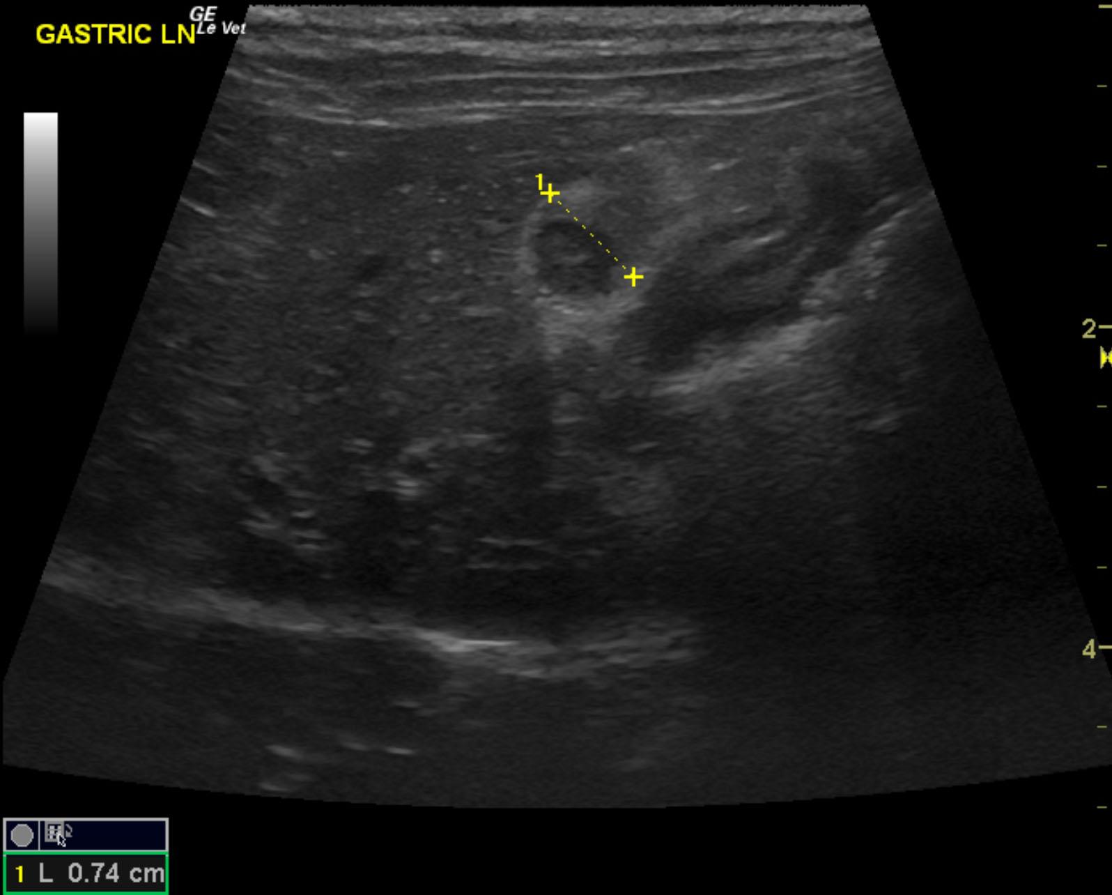
A 7-year-old SF DSH was presented for evaluation of a two-week period of vomiting food. Physical examination and blood work were within normal limits. There had been no response to fenbendazole therapy.
A 7-year-old SF DSH was presented for evaluation of a two-week period of vomiting food. Physical examination and blood work were within normal limits. There had been no response to fenbendazole therapy.