Jejunal foreign body in a 5 year old MN Boxer **Aug 2013 COM**

Case Study

Jejunal foreign body in a 5 year old MN Boxer **Aug 2013 COM**

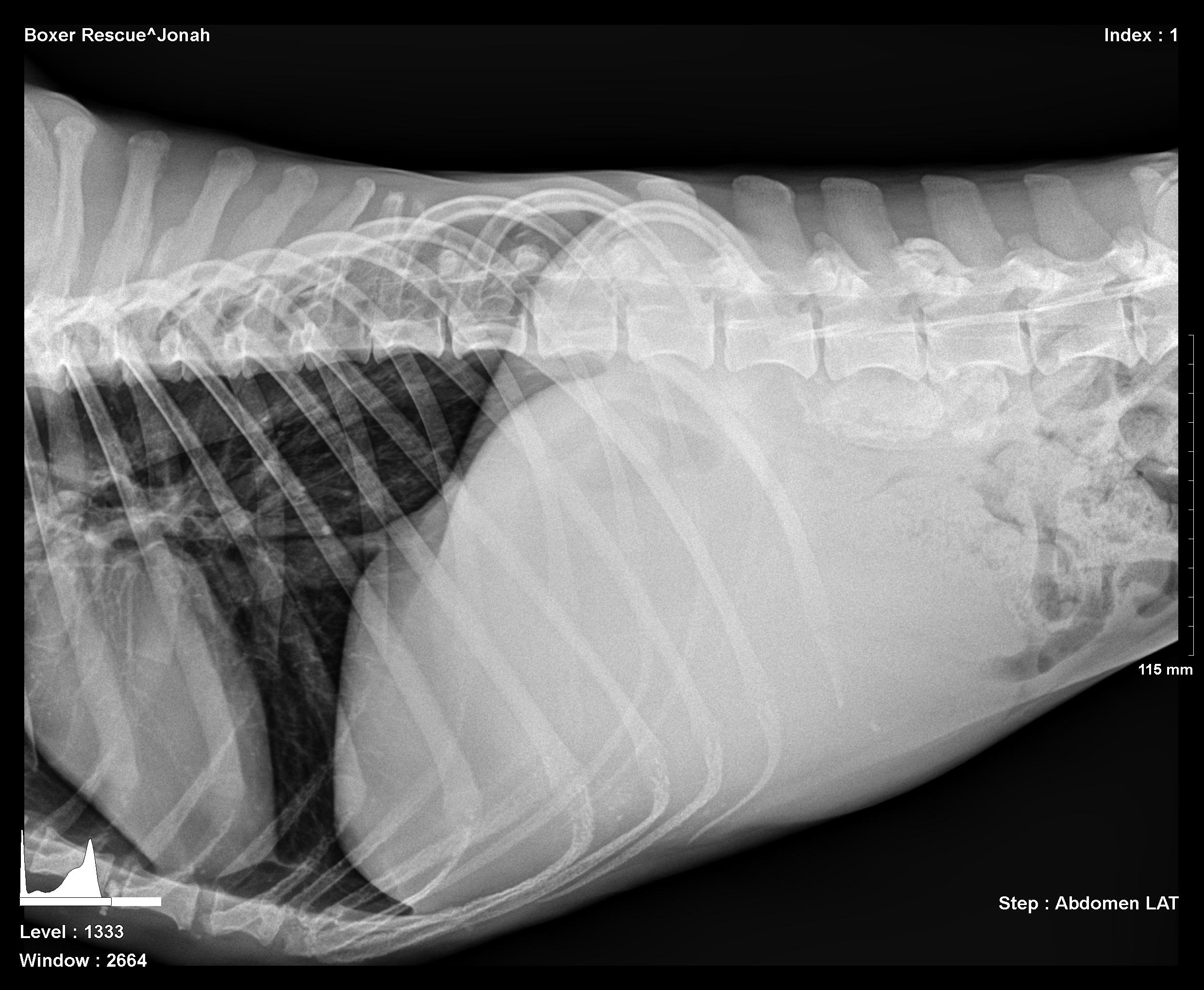
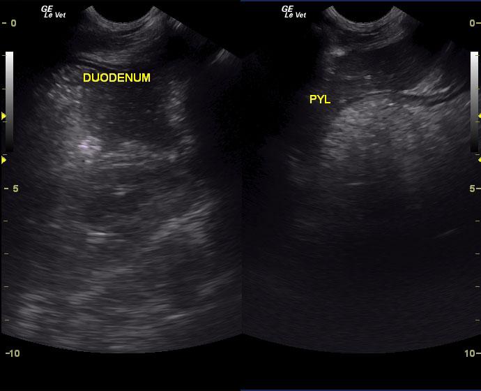
The patient is a 5 year old neutered male Boxer. The dog was presented to the clinic by a rescue organization. He was emaciated, vomiting, and anorexic. Bloodwork was unremarkable. Physical exam revealed a mid abdominal thickening potentially of intestinal origin. Lateral radiograph revealed a mid-cranial abdominal mass with mass effect upon the intestinal tract displacing the mesentery caudally. A volume-contracted heart was also visible (Image 1).

Sonographic Differential Diagnosis

Obstructive foreign body with likely underlying mural disease and minor regional omental inflammation.
Exploratory surgery with enterotomy, gastric and intestinal biopsies were recommended. Guarded prognosis depending on underlying histopathology whether concurrent intestinal neoplasia is present (versus inflammatory-related mural hypertrophy).

Image Interpretation

Upper GI stasis is evident in both the stomach and duodenum (image 2). A distinctly shadowing intestinal foreign body is seen (arrow) with mural thickening and loss of layering of the affected intestinal wall (Image 3). The 6 cm shadowing foreign body is visible with adjacent stasis followed by empty small intestine finalizing the obstructive pattern defined in our prior study (Video 1 and 2) (see: Sonographic Criteria for the Diagnosis of Gastrointestinal Obstruction in 39 Dogs and Cats, ECVIM 2009, http://www.sonopath.com/resources/articles). A minor amount of ill-defined surrounding fat is noted adjacent to the intestinal wall in the near field indicative of an active transmural disease process, which classifies this case as a surgical emergency before further breakdown of the intestinal wall occurs.

DX

Jejunal obstruction due to foreign body monkey toy

Outcome

The patient recovered well and is gaining weight readily (Image 8).

Comments

Special thanks to Dr. Harvey Hummel and Head Technician Diane McFadden & staff at Andover Animal Hospital, Andover, NJ, USA (http://www.andoveranimalhospital.com/) for their wonderful management and care for Jonah, the curious Boxer that just wanted to play with a monkey.


Clinical Differential Diagnosis

Neoplasia, intestinal obstruction, intestinal torsion, abdominal abscess or cyst, pancreatitis or pancreatic necrosis.

Sampling

Patient had exploratory surgery with solid foreign body removal (Image 4) that revealed an underlying monkey toy (plastic monkey from barrel of monkeys, image 5). Intestinal biopsy revealed fibrosing enteritis/peritonitis adjacent to the intestinal wall without evidence of neoplasia.

Patient Information

Patient Name : Jonah B
Gender : Male, Neutered
Species : Canine
Type of Imaging : Ultrasound
Status : Complete
Liz Wuz Here : Yes
Code : 04_00399

Clinical Signs

  • Anorexia
  • Vomiting
  • Weight loss

Exam Finding

  • Dehydration
  • Thickened Intestines
  • Weight loss

Images

image_1_jonahimage_2_jonahimage_3_jonahimage_6_jonahimage_7_jonah

Clinical Signs

  • Anorexia
  • Vomiting
  • Weight loss
Skip to content