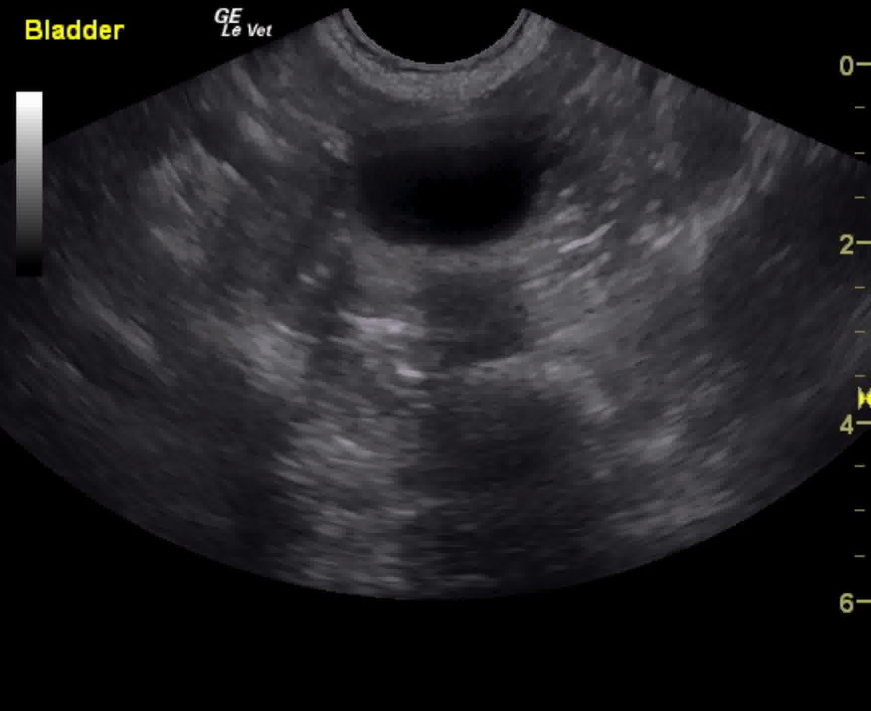
The urinary bladder presented minor mural thickening with slight, micropolypoid changes.
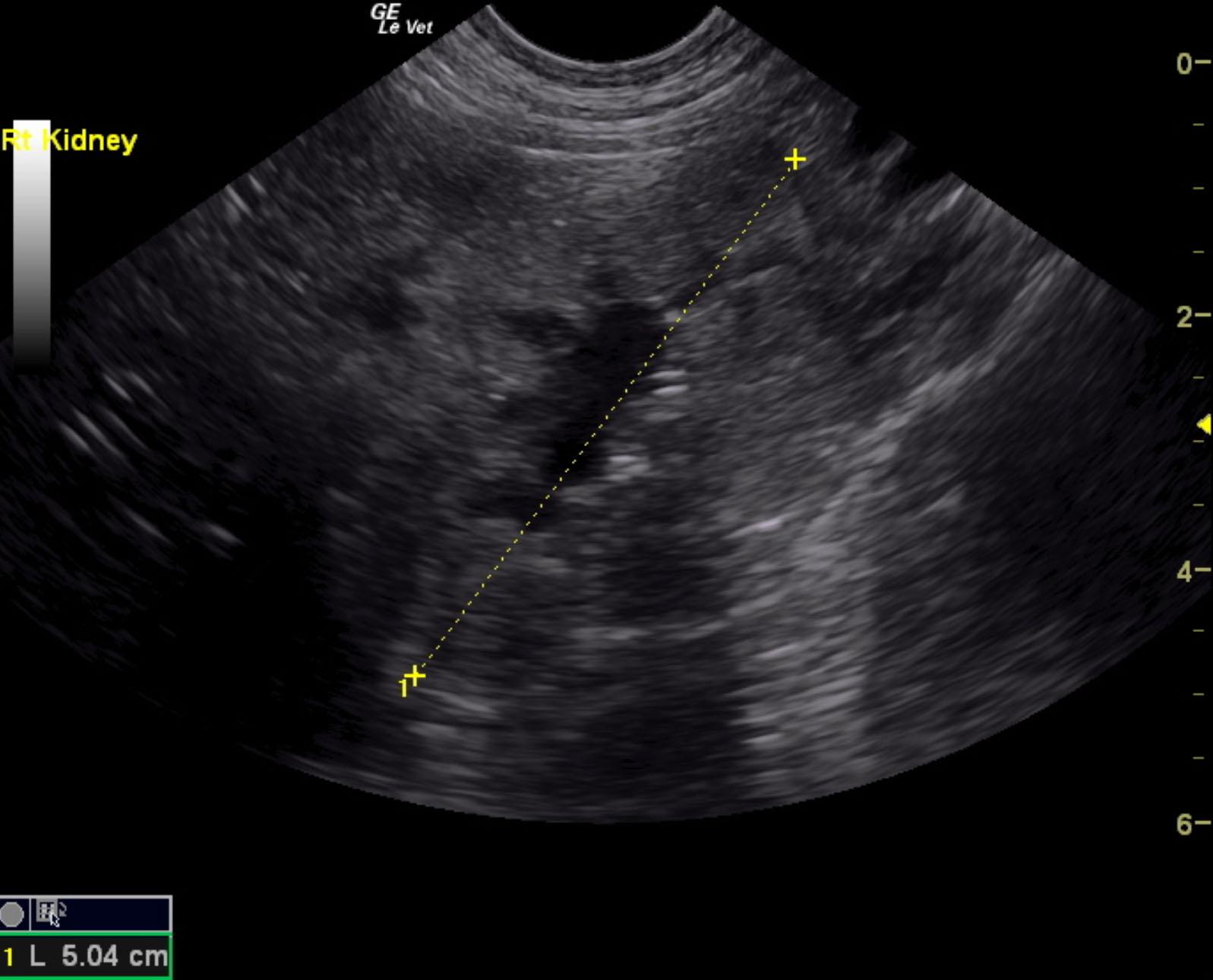
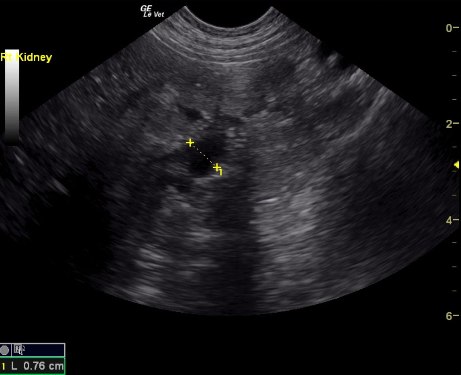
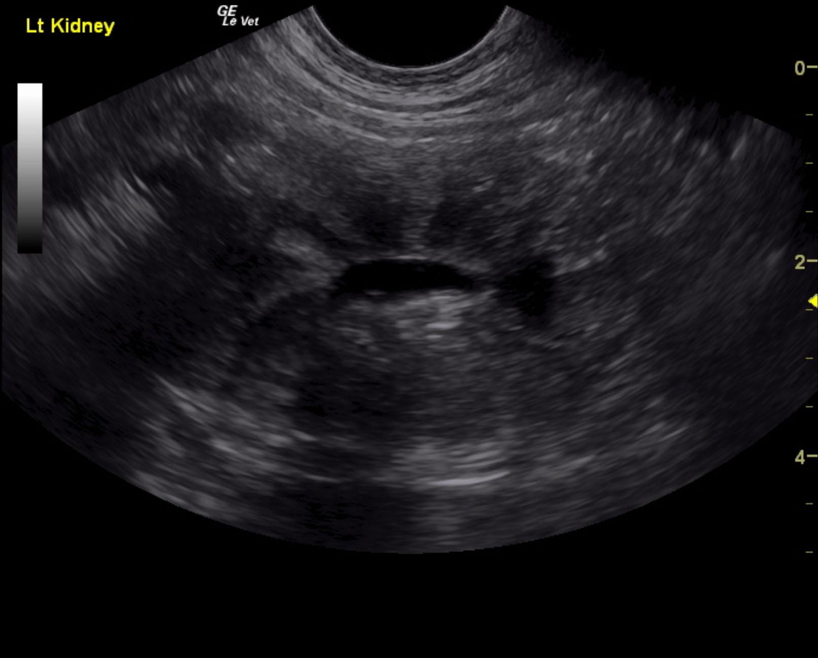
Both kidneys in this patient presented significant, dysplastic changes with cortical irregularity and disruption of the corticomedullary structure. A significant amount of microinfarctions and hyperechoic areas of mineralization and fibrosis as well as cortical cysts were noted. Color flow assessment of the renal cortices was significantly subnormal. This is consistent with chronic disease. The left kidney measured 5.08 cm. The right kidney presented pyelectasia that measured 0.76 cm. The right kidney measured 5.04 cm.