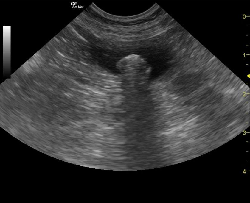
A 10-year-old MN Shih Tzu was presented for evaluation of intermittent stranguria and weight loss. On rectal palpation, there was some thickening of the prostate. Urinalysis and radiographs were both within normal limits and blood work showed elevated ALP activity.
A 10-year-old MN Shih Tzu was presented for evaluation of intermittent stranguria and weight loss. On rectal palpation, there was some thickening of the prostate. Urinalysis and radiographs were both within normal limits and blood work showed elevated ALP activity.